 Explore
 Validate
 Learn
Learn Western blot
 ELISA
ELISAAntibody data
- Antibody Data
- Antigen structure
- References [1]
- Comments [0]
- Validations
- Western blot [3]
- Immunocytochemistry [1]
Submit
Validation data
Reference
Comment
Report error
- Product number
- MA1-19426 - Provider product page

- Provider
- Invitrogen Antibodies
- Product name
- MAP2 Monoclonal Antibody (MT-08)
- Antibody type
- Monoclonal
- Antigen
- Other
- Description
- This antibody recognizes an epitope (aa 1375-1395) located in central domain of molecule Microtubule Associated Protein 2ab (MAP2ab), an intracellular antigen.
- Reactivity
- Human, Mouse, Porcine
- Host
- Mouse
- Isotype
- IgG
- Antibody clone number
- MT-08
- Vial size
- 100 µg
- Concentration
- 1 mg/mL
- Storage
- 4° C, do not freeze
Submitted references Global Landscape and Dynamics of Parkin and USP30-Dependent Ubiquitylomes in iNeurons during Mitophagic Signaling.
Ordureau A, Paulo JA, Zhang J, An H, Swatek KN, Cannon JR, Wan Q, Komander D, Harper JW
Molecular cell 2020 Mar 5;77(5):1124-1142.e10
Molecular cell 2020 Mar 5;77(5):1124-1142.e10
No comments: Submit comment
Supportive validation
- Submitted by
- Invitrogen Antibodies (provider)
- Main image
- Experimental details
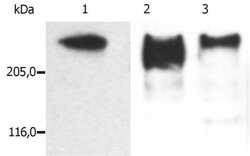
- Western blot analysis of microtubules partially purified from porcine brain lysate. Probed with MAP2 a, b monoclonal antibodies (Product # MA1-19166 (Lane 1)), (Product # MA1-19425 (Lane 2)), (Product # MA1-19426 (Lane 3)).

- Submitted by
- Invitrogen Antibodies (provider)
- Main image

- Experimental details
- Western blotting analysis (reducing conditions) of microtubules partially purified from porcine brain lysate. Lane 1: immunostaining with anti-MAP2 (MT-01); Lane 2: immunostaining with anti-MAP2 (MT-07); Lane 3: immunostaining with anti-MAP2 (MT-08) Monoclonal antibody (Product # MA1-19426).

- Submitted by
- Invitrogen Antibodies (provider)
- Main image
- Experimental details
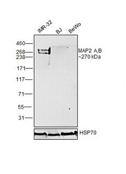
- Western blot was performed using Anti-MAP2 Monoclonal Antibody (MT-08) (Product # MA1-19426) and a ~270 kDa band corresponding to MAP2 A,B was observed in positive cell line IMR-32. Whole cell extracts (30 µg lysate) of IMR-32 (Lane 1), BJ (Lane 2) and BeWo (Lane 3) were electrophoresed using NuPAGE™ 3-8% Tris-Acetate Protein Gel (Product # EA0378BOX). Resolved proteins were then transferred onto a Nitrocellulose membrane (Product # IB23001) by iBlot® 2 Dry Blotting System (Product # IB21001). The blot was probed with the primary antibody (1:1,000 dilution) and detected by chemiluminescence with Goat anti-Mouse IgG (H+L) Superclonal™ Recombinant Secondary Antibody, HRP (Product # A28177, 1:4,000 dilution) using the iBright FL 1000 (Product # A32752). Chemiluminescent detection was performed using SuperSignal™ West Dura Extended Duration Substrate (Product # 34076).

Supportive validation
- Submitted by
- Invitrogen Antibodies (provider)
- Main image

- Experimental details
- Immunofluorescence analysis of Microtubule-associated protein 2 was performed using 70% confluent log phase SH-SY5Y cells differentiated to neurons (Retinoic acid 10 uM for 5 days). The cells were fixed with 4% paraformaldehyde for 10 minutes, permeabilized with 0.1% Triton™ X-100 for 15 minutes, and blocked with 2% BSA for 45 minutes at room temperature. The cells were labeled with MAP2 Monoclonal Antibody (MT-08) (Product # MA1-19426) at 1:100 dilution in 0.1% BSA, incubated at 4 degree celsius overnight and then labeled with Donkey anti-Mouse IgG (H+L) Highly Cross-Adsorbed Secondary Antibody, Alexa Fluor Plus 488 (Product # A32766), (1:2000 dilution), for 45 minutes at room temperature (Panel a: Green). Nuclei (Panel b: Blue) were stained with ProLong™ Diamond Antifade Mountant with DAPI (Product # P36962). F-actin (Panel c: Red) was stained with Rhodamine Phalloidin (Product # R415, 1:300 dilution). Panel d represents the merged image showing neuronal dendrites, cytoplasm, plasma membrane and cytoskeleton localization. Panel e represents undifferentiated SH-SY5Y cells. Panel f represents control cells with no primary antibody to assess background. The images were captured at 60X magnification.
