Explore
 Validate
 Learn
Learn Western blot
 Immunocytochemistry
Antibody data
- Antibody Data
- Antigen structure
- References [6]
- Comments [0]
- Validations
- Immunocytochemistry [1]
- Immunohistochemistry [1]
Submit
Validation data
Reference
Comment
Report error
- Product number
- HPA012828 - Provider product page

- Provider
- Atlas Antibodies
- Proper citation
- Atlas Antibodies Cat#HPA012828, RRID:AB_1853946
- Product name
- Anti-MAP2
- Antibody type
- Polyclonal
- Description
- Polyclonal Antibody against Human MAP2, Gene description: microtubule-associated protein 2, Alternative Gene Names: MAP2A, MAP2B, MAP2C, Validated applications: ICC, IHC, WB, Uniprot ID: P11137, Storage: Store at +4°C for short term storage. Long time storage is recommended at -20°C.
- Reactivity
- Human
- Host
- Rabbit
- Conjugate
- Unconjugated
- Isotype
- IgG
- Vial size
- 100 µl
- Concentration
- 0.05 mg/ml
- Storage
- Store at +4°C for short term storage. Long time storage is recommended at -20°C.
- Handling
- The antibody solution should be gently mixed before use.
Submitted references Ras Inhibitor Lonafarnib Rescues Structural and Functional Impairments of Synapses of Aβ1-42 Mice via α7nAChR-Dependent BDNF Upregulation
Carbon Nanotube–Hydrogel Composites Facilitate Neuronal Differentiation While Maintaining Homeostasis of Network Activity
Controlling the Switch from Neurogenesis to Pluripotency during Marmoset Monkey Somatic Cell Reprogramming with Self-Replicating mRNAs and Small Molecules
α‑synuclein induces apoptosis of astrocytes by causing dysfunction of the endoplasmic reticulum‑Golgi compartment
Antibodies biotinylated using a synthetic Z-domain from protein A provide stringent in situ protein detection.
The Human Protein Atlas—a tool for pathology
Cai C, Wang L, Li S, Lou S, Luo J, Fu D, Chen T
The Journal of Neuroscience 2022;42(31):6090-6107
The Journal of Neuroscience 2022;42(31):6090-6107
Carbon Nanotube–Hydrogel Composites Facilitate Neuronal Differentiation While Maintaining Homeostasis of Network Activity
Ye L, Ji H, Liu J, Tu C, Kappl M, Koynov K, Vogt J, Butt H
Advanced Materials 2021;33(41)
Advanced Materials 2021;33(41)
Controlling the Switch from Neurogenesis to Pluripotency during Marmoset Monkey Somatic Cell Reprogramming with Self-Replicating mRNAs and Small Molecules
Petkov S, Dressel R, Rodriguez-Polo I, Behr R
Cells 2020;9(11):2422
Cells 2020;9(11):2422
α‑synuclein induces apoptosis of astrocytes by causing dysfunction of the endoplasmic reticulum‑Golgi compartment
Liu M, Qin L, Wang L, Tan J, Zhang H, Tang J, Shen X, Tan L, Wang C
Molecular Medicine Reports 2018
Molecular Medicine Reports 2018
Antibodies biotinylated using a synthetic Z-domain from protein A provide stringent in situ protein detection.
Andersson S, Konrad A, Ashok N, Pontén F, Hober S, Asplund A
The journal of histochemistry and cytochemistry : official journal of the Histochemistry Society 2013 Nov;61(11):773-84
The journal of histochemistry and cytochemistry : official journal of the Histochemistry Society 2013 Nov;61(11):773-84
The Human Protein Atlas—a tool for pathology
Pontén F, Jirström K, Uhlen M
The Journal of Pathology 2008;216(4):387-393
The Journal of Pathology 2008;216(4):387-393
No comments: Submit comment
Supportive validation
- Submitted by
- Atlas Antibodies (provider)
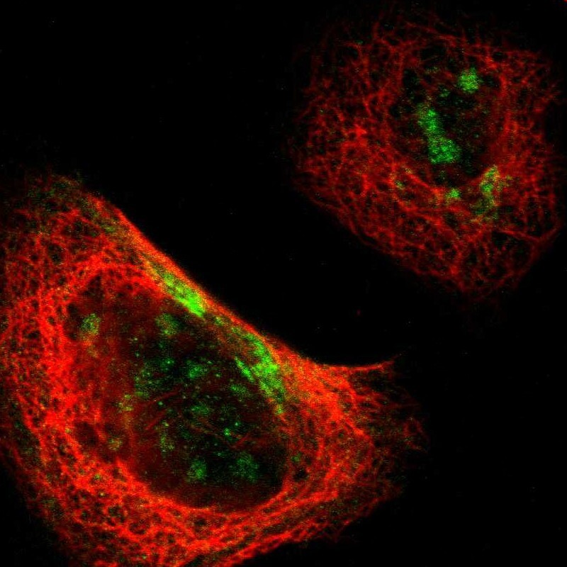
- Main image

- Experimental details
- Immunofluorescent staining of human cell line A-431 shows localization to nucleoli & cytosol.
- Sample type
- Human
Supportive validation
- Submitted by
- Atlas Antibodies (provider)
- Enhanced method
- Orthogonal validation
- Main image

- Experimental details
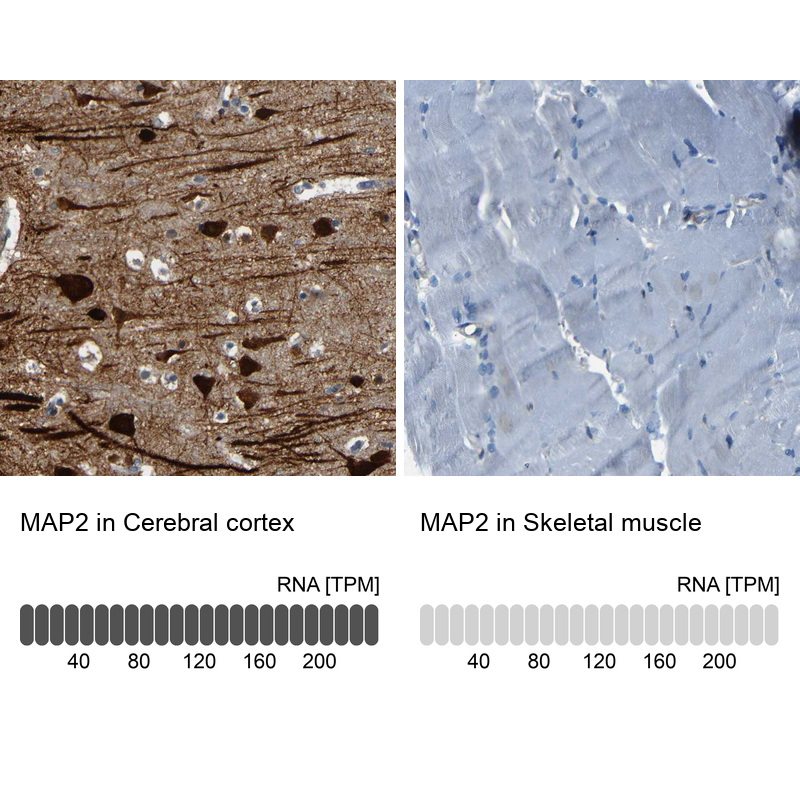
- Immunohistochemistry analysis in human cerebral cortex and skeletal muscle tissues using HPA012828 antibody. Corresponding MAP2 RNA-seq data are presented for the same tissues.
- Sample type
- Human
- Protocol
- Protocol