Explore
 Validate
 Learn
Learn Western blot
 Immunocytochemistry
 Immunoprecipitation
Antibody data
- Antibody Data
- Antigen structure
- References [2]
- Comments [0]
- Validations
- Immunocytochemistry [2]
- Other assay [3]
Submit
Validation data
Reference
Comment
Report error
- Product number
- 37-2100 - Provider product page

- Provider
- Invitrogen Antibodies
- Product name
- PEBP1 Monoclonal Antibody (3E12D7)
- Antibody type
- Monoclonal
- Antigen
- Other
- Reactivity
- Human, Mouse
- Host
- Mouse
- Isotype
- IgG
- Antibody clone number
- 3E12D7
- Vial size
- 100 μg
- Concentration
- 0.5 mg/mL
- Storage
- -20°C
Submitted references RKIP Regulates Differentiation-Related Features in Melanocytic Cells.
Proteomic characterization of differentially expressed proteins in breast cancer: Expression of hnRNP H1, RKIP and GRP78 is strongly associated with HER-2/neu status.
Penas C, Apraiz A, Muñoa I, Arroyo-Berdugo Y, Rasero J, Ezkurra PA, Velasco V, Subiran N, Bosserhoff AK, Alonso S, Asumendi A, Boyano MD
Cancers 2020 Jun 3;12(6)
Cancers 2020 Jun 3;12(6)
Proteomic characterization of differentially expressed proteins in breast cancer: Expression of hnRNP H1, RKIP and GRP78 is strongly associated with HER-2/neu status.
Zhang D, Tai LK, Wong LL, Putti TC, Sethi SK, Teh M, Koay ES
Proteomics. Clinical applications 2008 Jan;2(1):99-107
Proteomics. Clinical applications 2008 Jan;2(1):99-107
No comments: Submit comment
Supportive validation
- Submitted by
- Invitrogen Antibodies (provider)
- Main image
- Experimental details
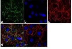
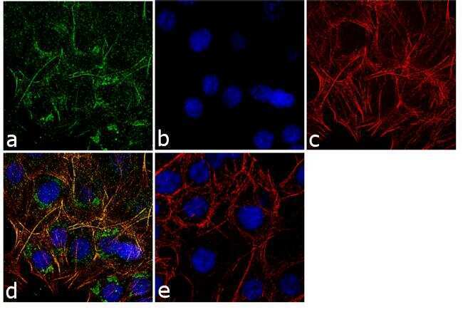
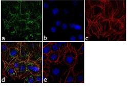
- Immunofluorescence analysis of PEBP1 was performed using 70% confluent log phase HCT-116 cells. The cells were fixed with 4% paraformaldehyde for 10 minutes, permeabilized with 0.1% Triton™ X-100 for 10 minutes, and blocked with 1% BSA for 1 hour at room temperature. The cells were labeled with Prostate Specific Acid Phosphatase (PASE/4LJ) Mouse Monoclonal Antibody (3721) at 2 µg/mL in 0.1% BSA and incubated for 3 hours at room temperature and then labeled with Goat anti-Mouse IgG (H+L) Superclonal™ Secondary Antibody, Alexa Fluor® 488 conjugate (Product # A28175) at a dilution of 1:2000 for 45 minutes at room temperature (Panel a: green). Nuclei (Panel b: blue) were stained with SlowFade® Gold Antifade Mountant with DAPI (Product # S36938). F-actin (Panel c: red) was stained with Alexa Fluor® 555 Rhodamine Phalloidin (Product # R415, 1:300). Panel d represents the merged image showing cytoplasmic localization. Panel e shows the control without primary antibody. The images were captured at 60X magnification.
- Submitted by
- Invitrogen Antibodies (provider)
- Main image
- Experimental details
- Immunofluorescence analysis of PEBP1 was performed using 70% confluent log phase HCT-116 cells. The cells were fixed with 4% paraformaldehyde for 10 minutes, permeabilized with 0.1% Triton™ X-100 for 10 minutes, and blocked with 1% BSA for 1 hour at room temperature. The cells were labeled with Prostate Specific Acid Phosphatase (PASE/4LJ) Mouse Monoclonal Antibody (3721) at 2 µg/mL in 0.1% BSA and incubated for 3 hours at room temperature and then labeled with Goat anti-Mouse IgG (H+L) Superclonal™ Secondary Antibody, Alexa Fluor® 488 conjugate (Product # A28175) at a dilution of 1:2000 for 45 minutes at room temperature (Panel a: green). Nuclei (Panel b: blue) were stained with SlowFade® Gold Antifade Mountant with DAPI (Product # S36938). F-actin (Panel c: red) was stained with Alexa Fluor® 555 Rhodamine Phalloidin (Product # R415, 1:300). Panel d represents the merged image showing cytoplasmic localization. Panel e shows the control without primary antibody. The images were captured at 60X magnification.

Supportive validation
- Submitted by
- Invitrogen Antibodies (provider)
- Main image
- Experimental details
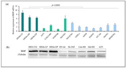
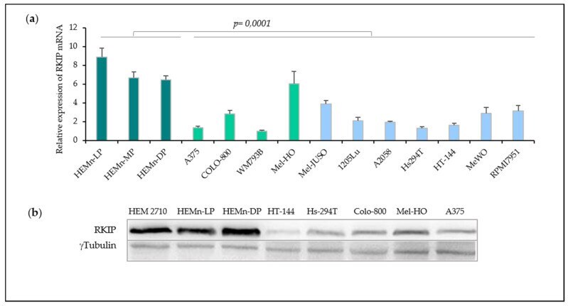
- Figure 2 RKIP expression in cell culture of primary melanocytes and melanomas. ( a ) RKIP mRNA expression in primary melanocytes and melanoma cell lines. RNA from three human melanocytes (dark green) lightly (HEMn-LP), moderately (HEMn-MP), and darkly (HEMn-DP) pigmented neonatal foreskin lines together with four primary melanomas cell lines (light green) and seven metastatic melanomas cell lines (light blue) were analyzed by RT-qPCR. ( b ) RKIP protein expression in the three melanocytes cell lines and five melanoma cell lines assessed by Western Blot. Tubulin expression was used as loading control. Figure is representative of three independent experiments.
- Submitted by
- Invitrogen Antibodies (provider)
- Main image

- Experimental details
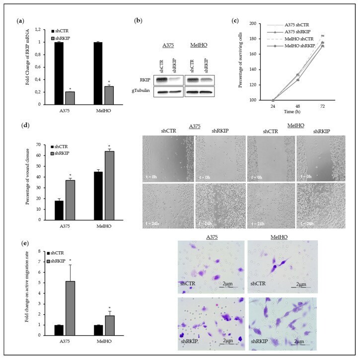
- Figure 3 Modulation of RKIP expression in primary melanoma cell lines. ( a ) RKIP mRNA levels in RKIP-downregulated A375 and MelHO primary melanoma cell lines. A375 and MelHO cells were transduced with RKIP shRNA Lentiviral Particles or Control shRNA Lentiviral Particles following the manufacturer's instruction. Two days after infection, the cells were selected with Puromycin to get stable cell lines; ( b ) Western Blot assay showed the RKIP-downregulation in A375 and MelHO melanoma cells; ( c ) Proliferation rate in A375 and MelHO primary melanomas after RKIP downregulation. The viability of control melanoma cells (with an empty vector) and stable RKIP transfected clones were subjected to XTT assays for 24, 48, and 72 h. Results of each experiment are expressed related to the values obtained for the transfection control. Data is given as a mean +- SD of at least three experiments of different transfection; ( d ) Fold change on wound healing rate in A375 and MelHO primary melanoma after RKIP downregulation; ( e ) Fold change on active migration rate in presence of collagen in primary melanoma after RKIP downregulation. The histograms in ( d ) and ( e ) show the average of three independent assays with six replicates per assay and representative pictures have been included. * p -value < 0.05.
- Submitted by
- Invitrogen Antibodies (provider)
- Main image
- Experimental details
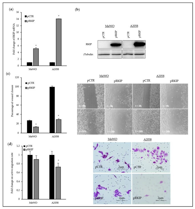
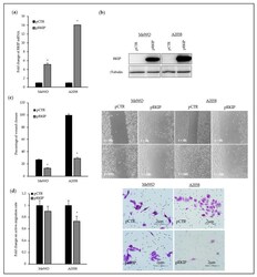
- Figure 4 Modulation of RKIP expression in MeWO and A2058 metastatic melanoma cell lines. ( a ) RKIP mRNA levels in RKIP-upregulated MeWO and A2058 metastatic melanoma cell lines. A2058 and MeWO cell lines were transfected with overexpressing plasmid for RKIP (pRKIP) or empty plasmid (pCTR) using Lipofectamine 2000 as transfection agent. All of the transfection experiments were performed with 500 ng of DNA. ( b ) Western Blot assay showed the RKIP-upregulation in MeWO and A2058 melanoma cells; ( c ) Fold change on wound healing rate in MeWO and A2058 metastatic melanoma after RKIP upregulation. The experimental assays were performed at least after 24 h of RKIP transfection; ( d ) Fold change on active migration rate in presence of collagen in metastatic melanoma after RKIP upregulation. The histograms in ( c ) and ( d ) show the average of three independent assays with six replicates per assay and representative pictures have been included. * p -value < 0.05.