 Explore
 Validate
 Learn
Learn Western blot
Antibody data
- Antibody Data
- Antigen structure
- References [1]
- Comments [0]
- Validations
- Western blot [2]
Submit
Validation data
Reference
Comment
Report error
- Product number
- AF5234 - Provider product page

- Provider
- R&D Systems
- Product name
- Human CRTAC1 Isoform 1 Antibody
- Antibody type
- Polyclonal
- Description
- Immunogen affinity purified. Detects human CRTAC1 Isoform 1 in direct ELISAs and Western blots. In Western blots, approximately 10% cross-reactivity with recombinant human CRTAC1 Isoform 2 is observed.
- Reactivity
- Human
- Host
- Sheep
- Conjugate
- Unconjugated
- Antigen sequence
Q9NQ79- Isotype
- IgG
- Vial size
- 100 ug
- Concentration
- LYOPH
- Storage
- Use a manual defrost freezer and avoid repeated freeze-thaw cycles. 12 months from date of receipt, -20 to -70 °C as supplied. 1 month, 2 to 8 °C under sterile conditions after reconstitution. 6 months, -20 to -70 °C under sterile conditions after reconstitution.
Submitted references Cerebrospinal fluid level of Nogo receptor 1 antagonist lateral olfactory tract usher substance (LOTUS) correlates inversely with the extent of neuroinflammation.
Takahashi K, Takeuchi H, Kurihara Y, Doi H, Kunii M, Tanaka K, Nakamura H, Fukai R, Tomita-Katsumoto A, Tada M, Higashiyama Y, Joki H, Koyano S, Takei K, Tanaka F
Journal of neuroinflammation 2018 Feb 17;15(1):46
Journal of neuroinflammation 2018 Feb 17;15(1):46
No comments: Submit comment
Supportive validation
- Submitted by
- R&D Systems (provider)
- Main image
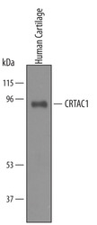
- Experimental details
- Detection of Human CRTAC1 by Western Blot. Western blot shows lysates of human cartilage tissue. PVDF membrane was probed with 1 µg/mL of Sheep Anti-Human CRTAC1 Isoform 1 Antigen Affinity-purified Polyclonal Antibody (Catalog # AF5234) followed by HRP-conjugated Anti-Sheep IgG Secondary Antibody (Catalog # HAF016). A specific band was detected for CRTAC1 at approximately 95 kDa (as indicated). This experiment was conducted under reducing conditions and using Immunoblot Buffer Group 8.

- Submitted by
- R&D Systems (provider)
- Main image
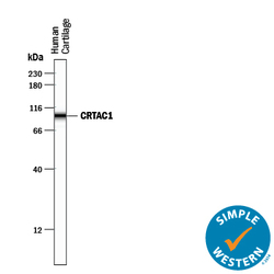
- Experimental details
- Detection of Human CRTAC1 by Simple WesternTM. Simple Western lane view shows lysates of human cartilage tissue, loaded at 0.5 mg/mL. A specific band was detected for CRTAC1 at approximately 97 kDa (as indicated) using 10 µg/mL of Sheep Anti-Human CRTAC1 Isoform 1 Antigen Affinity-purified Polyclonal Antibody (Catalog # AF5234) followed by 1:50 dilution of HRP-conjugated Anti-Sheep IgG Secondary Antibody (Catalog # HAF016). This experiment was conducted under reducing conditions and using the 12-230 kDa separation system.
