Explore
 Validate
 Learn
Learn Western blot
Antibody data
- Antibody Data
- Antigen structure
- References [4]
- Comments [0]
- Validations
- Western blot [1]
- Immunocytochemistry [4]
- Immunohistochemistry [3]
- Flow cytometry [5]
- Other assay [2]
Submit
Validation data
Reference
Comment
Report error
- Product number
- MA3-062 - Provider product page

- Provider
- Invitrogen Antibodies
- Product name
- SPTBN1 Monoclonal Antibody (4C3)
- Antibody type
- Monoclonal
- Antigen
- Purifed from natural sources
- Description
- MA3-062 detects spectrin from mouse, rat and human erythrocytes, brain and muscle cells. This antibody has been shown to specifically detect the two known alternatively spliced forms of spectrin, beta-1 epsilon-1, present in erythrocytes, and beta-1 epsilon-2, present in nerve and striated muscle cells. It does not cross-react with alpha-2 spectrin or either of the fodrin subunits. MA3-062 has been successfully used in Western blot, immunohistochemistry (paraffin) and immunofluorescence procedures. By Western blot, this antibody detects a ~246 kDa protein representing beta-1 spectrin in rat skeletal muscle homogenate. Immunofluorescence staining of beta-1 spectrin in rat skeletal muscle fibers with MA3-062 results in intense staining of the sarcolemma. In immunofluorescence, antigen integrity can be compromised if aldehyde fixatives are left in contact with the protein for extended periods of time. If paraformaldehyde is used as a fixative, exposure should be limited to 5 minutes or less of no more than a 2% solution. The MA3-062 antigen is purified human erythrocyte beta-1 spectrin.
- Reactivity
- Human, Mouse, Rat
- Host
- Mouse
- Isotype
- IgG
- Antibody clone number
- 4C3
- Vial size
- 100 μL
- Concentration
- Conc. Not Determined
- Storage
- -20°C, Avoid Freeze/Thaw Cycles
Submitted references MEF2c-Dependent Downregulation of Myocilin Mediates Cancer-Induced Muscle Wasting and Associates with Cachexia in Patients with Cancer.
Co-expression analysis of pancreatic cancer proteome reveals biology and prognostic biomarkers.
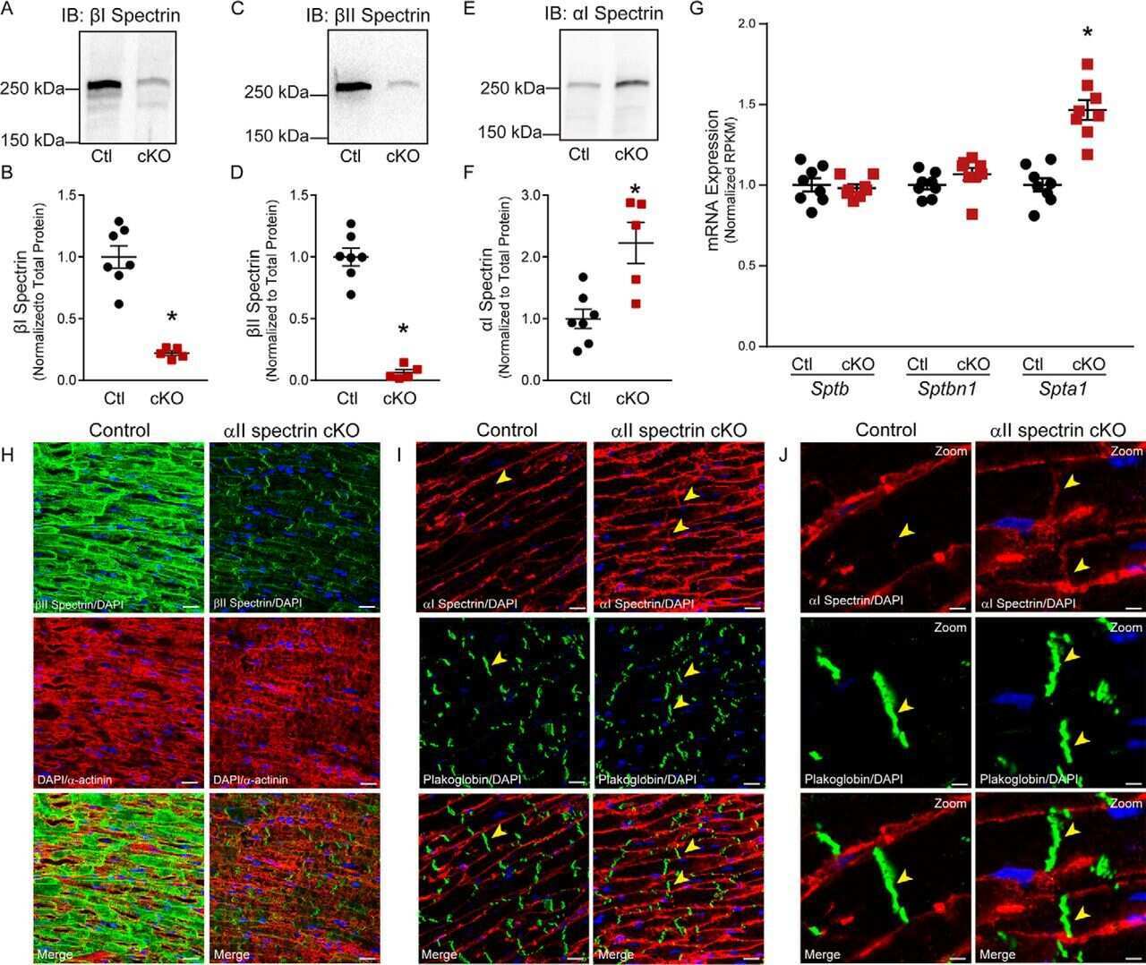
Defining new mechanistic roles for αII spectrin in cardiac function.
Two populations of beta-spectrin in rat skeletal muscle.
Judge SM, Deyhle MR, Neyroud D, Nosacka RL, D'Lugos AC, Cameron ME, Vohra RS, Smuder AJ, Roberts BM, Callaway CS, Underwood PW, Chrzanowski SM, Batra A, Murphy ME, Heaven JD, Walter GA, Trevino JG, Judge AR
Cancer research 2020 May 1;80(9):1861-1874
Cancer research 2020 May 1;80(9):1861-1874
Co-expression analysis of pancreatic cancer proteome reveals biology and prognostic biomarkers.
Mantini G, Vallés AM, Le Large TYS, Capula M, Funel N, Pham TV, Piersma SR, Kazemier G, Bijlsma MF, Giovannetti E, Jimenez CR
Cellular oncology (Dordrecht) 2020 Dec;43(6):1147-1159
Cellular oncology (Dordrecht) 2020 Dec;43(6):1147-1159
Defining new mechanistic roles for αII spectrin in cardiac function.
Lubbers ER, Murphy NP, Musa H, Huang CY, Gupta R, Price MV, Han M, Daoud E, Gratz D, El Refaey M, Xu X, Hoeflinger NK, Friel EL, Lancione P, Wallace MJ, Cavus O, Simmons SL, Williams JL, Skaf M, Koenig SN, Janssen PML, Rasband MN, Hund TJ, Mohler PJ
The Journal of biological chemistry 2019 Jun 14;294(24):9576-9591
The Journal of biological chemistry 2019 Jun 14;294(24):9576-9591
Two populations of beta-spectrin in rat skeletal muscle.
Porter GA, Scher MG, Resneck WG, Porter NC, Fowler VM, Bloch RJ
Cell motility and the cytoskeleton 1997;37(1):7-19
Cell motility and the cytoskeleton 1997;37(1):7-19
No comments: Submit comment
Supportive validation
- Submitted by
- Invitrogen Antibodies (provider)
- Main image
- Experimental details
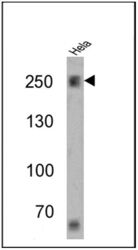
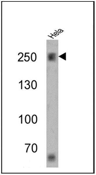
- Western blot analysis of Spectrin beta-1 was performed by loading 25 µg of Hela cell lysates onto an SDS polyacrylamide gel. Proteins were transferred to a PVDF membrane and blocked at 4ºC overnight. The membrane was probed with a Spectrin beta-1 monoclonal antibody (Product # MA3-062) at a dilution of 1:1000 overnight at 4°C, washed in TBST, and probed with an HRP-conjugated secondary antibody for 1 hr at room temperature in the dark. Chemiluminescent detection was performed using Pierce ECL Plus Western Blotting Substrate (Product # 32132). Results show a band at ~246 kDa.
Supportive validation
- Submitted by
- Invitrogen Antibodies (provider)
- Main image
- Experimental details
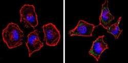
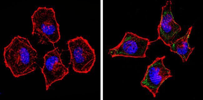
- Immunofluorescent analysis of Spectrin beta-1 (green) showing staining in the cytoplasm of Hela cells (right) compared to a negative control without primary antibody (left). Formalin-fixed cells were permeabilized with 0.1% Triton X-100 in TBS for 5-10 minutes and blocked with 3% BSA-PBS for 30 minutes at room temperature. Cells were probed with a Spectrin beta-1 monoclonal antibody (Product # MA3-062) in 3% BSA-PBS at a dilution of 1:100 and incubated overnight at 4 ºC in a humidified chamber. Cells were washed with PBST and incubated with a DyLight-conjugated secondary antibody in PBS at room temperature in the dark. F-actin (red) was stained with a fluorescent red phalloidin and nuclei (blue) were stained with Hoechst or DAPI. Images were taken at a magnification of 60x.

- Submitted by
- Invitrogen Antibodies (provider)
- Main image

- Experimental details
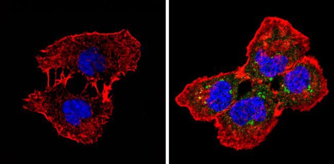
- Immunofluorescent analysis of Spectrin beta-1 (green) showing staining in the cytoplasm of A431 cells (right) compared to a negative control without primary antibody (left). Formalin-fixed cells were permeabilized with 0.1% Triton X-100 in TBS for 5-10 minutes and blocked with 3% BSA-PBS for 30 minutes at room temperature. Cells were probed with a Spectrin beta-1 monoclonal antibody (Product # MA3-062) in 3% BSA-PBS at a dilution of 1:20 and incubated overnight at 4 ºC in a humidified chamber. Cells were washed with PBST and incubated with a DyLight-conjugated secondary antibody in PBS at room temperature in the dark. F-actin (red) was stained with a fluorescent red phalloidin and nuclei (blue) were stained with Hoechst or DAPI. Images were taken at a magnification of 60x.

- Submitted by
- Invitrogen Antibodies (provider)
- Main image

- Experimental details
- Immunofluorescent analysis of Spectrin beta-1 (green) showing staining in the cytoplasm of Hela cells (right) compared to a negative control without primary antibody (left). Formalin-fixed cells were permeabilized with 0.1% Triton X-100 in TBS for 5-10 minutes and blocked with 3% BSA-PBS for 30 minutes at room temperature. Cells were probed with a Spectrin beta-1 monoclonal antibody (Product # MA3-062) in 3% BSA-PBS at a dilution of 1:100 and incubated overnight at 4 ºC in a humidified chamber. Cells were washed with PBST and incubated with a DyLight-conjugated secondary antibody in PBS at room temperature in the dark. F-actin (red) was stained with a fluorescent red phalloidin and nuclei (blue) were stained with Hoechst or DAPI. Images were taken at a magnification of 60x.
- Submitted by
- Invitrogen Antibodies (provider)
- Main image
- Experimental details
- Immunofluorescent analysis of Spectrin beta-1 (green) showing staining in the cytoplasm of A431 cells (right) compared to a negative control without primary antibody (left). Formalin-fixed cells were permeabilized with 0.1% Triton X-100 in TBS for 5-10 minutes and blocked with 3% BSA-PBS for 30 minutes at room temperature. Cells were probed with a Spectrin beta-1 monoclonal antibody (Product # MA3-062) in 3% BSA-PBS at a dilution of 1:20 and incubated overnight at 4 ºC in a humidified chamber. Cells were washed with PBST and incubated with a DyLight-conjugated secondary antibody in PBS at room temperature in the dark. F-actin (red) was stained with a fluorescent red phalloidin and nuclei (blue) were stained with Hoechst or DAPI. Images were taken at a magnification of 60x.
Supportive validation
- Submitted by
- Invitrogen Antibodies (provider)
- Main image
- Experimental details
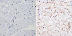
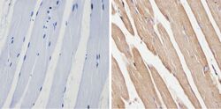
- Immunohistochemistry analysis of Spectrin beta-1 showing positive staining in the membrane of paraffin-treated Human skeletal muscle (right) compared with a negative control in the absence of primary antibody (left). To expose target proteins, antigen retrieval method was performed using 10mM sodium citrate (pH 6.0) microwaved for 8-15 min. Following antigen retrieval, tissues were blocked in 3% H2O2-methanol for 15 min at room temperature, washed with ddH2O and PBS, and then probed with a Spectrin beta-1 monoclonal antibody (Product # MA3-062) diluted by 3% BSA-PBS at a dilution of 1:200 overnight at 4°C in a humidified chamber. Tissues were washed extensively PBST and detection was performed using an HRP-conjugated secondary antibody followed by colorimetric detection using a DAB kit. Tissues were counterstained with hematoxylin and dehydrated with ethanol and xylene to prep for mounting.

- Submitted by
- Invitrogen Antibodies (provider)
- Main image
- Experimental details
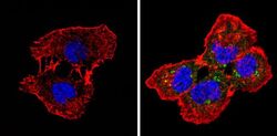
- Immunohistochemistry analysis of Spectrin beta-1 showing positive staining in the cytoplasm and membrane of paraffin-treated Mouse skeletal muscle (right) compared with a negative control in the absence of primary antibody (left). To expose target proteins, antigen retrieval method was performed using 10mM sodium citrate (pH 6.0) microwaved for 8-15 min. Following antigen retrieval, tissues were blocked in 3% H2O2-methanol for 15 min at room temperature, washed with ddH2O and PBS, and then probed with a Spectrin beta-1 monoclonal antibody (Product # MA3-062) diluted by 3% BSA-PBS at a dilution of 1:20 overnight at 4°C in a humidified chamber. Tissues were washed extensively PBST and detection was performed using an HRP-conjugated secondary antibody followed by colorimetric detection using a DAB kit. Tissues were counterstained with hematoxylin and dehydrated with ethanol and xylene to prep for mounting.

- Submitted by
- Invitrogen Antibodies (provider)
- Main image
- Experimental details
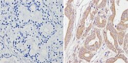
- Immunohistochemistry analysis of Spectrin beta-1 showing positive staining in the cytoplasm of paraffin-treated Human prostate carcinoma (right) compared with a negative control in the absence of primary antibody (left). To expose target proteins, antigen retrieval method was performed using 10mM sodium citrate (pH 6.0) microwaved for 8-15 min. Following antigen retrieval, tissues were blocked in 3% H2O2-methanol for 15 min at room temperature, washed with ddH2O and PBS, and then probed with a Spectrin beta-1 monoclonal antibody (Product # MA3-062) diluted by 3% BSA-PBS at a dilution of 1:20 overnight at 4°C in a humidified chamber. Tissues were washed extensively PBST and detection was performed using an HRP-conjugated secondary antibody followed by colorimetric detection using a DAB kit. Tissues were counterstained with hematoxylin and dehydrated with ethanol and xylene to prep for mounting.

Supportive validation
- Submitted by
- Invitrogen Antibodies (provider)
- Main image
- Experimental details
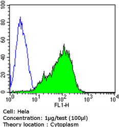
- Flow cytometry analysis of Spectrin beta-1 in Hela cells compared to an isotype control (blue). Cells were harvested, adjusted to a concentration of 1-5x10^6 cells/mL, fixed with 2% paraformaldehyde and washed with PBS. Cells were penetrated by dropping the supernatant, adding 90% methanol and incubated for 10 minutes at room temperature. Follwing penetration, cells were blocked with a 2% solution of BSA-PBS for 30 min at room temperature and incubated with a Spectrin beta-1 monoclonal antibody (Product # MA3-062) at a dilution of 1 µg/test for 60 min at room temperature. Cells were then incubated for 40 min at room temperature in the dark using a Dylight 488-conjugated goat anti-mouse IgG (H+L) secondary antibody and re-suspended in PBS for FACS analysis.

- Submitted by
- Invitrogen Antibodies (provider)
- Main image

- Experimental details
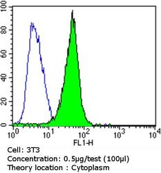
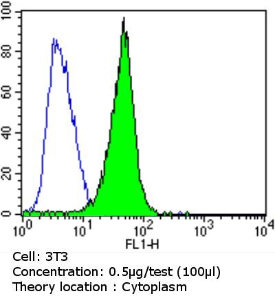
- Flow cytometry analysis of Spectrin beta-1 in NIH/3T3 cells compared to an isotype control (blue). Cells were harvested, adjusted to a concentration of 1-5x10^6 cells/mL, fixed with 2% paraformaldehyde and washed with PBS. Cells were penetrated by dropping the supernatant, adding 90% methanol and incubated for 10 minutes at room temperature. Follwing penetration, cells were blocked with a 2% solution of BSA-PBS for 30 min at room temperature and incubated with a Spectrin beta-1 monoclonal antibody (Product # MA3-062) at a dilution of 0.5 µg/test for 60 min at room temperature. Cells were then incubated for 40 min at room temperature in the dark using a Dylight 488-conjugated goat anti-mouse IgG (H+L) secondary antibody and re-suspended in PBS for FACS analysis.

- Submitted by
- Invitrogen Antibodies (provider)
- Main image
- Experimental details
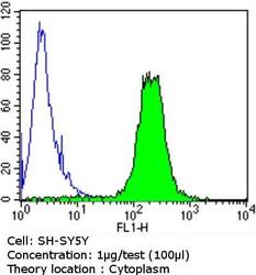
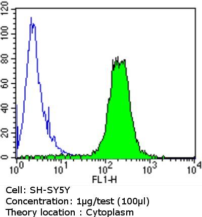
- Flow cytometry analysis of Spectrin beta-1 in SH-SY5Y cells compared to an isotype control (blue). Cells were harvested, adjusted to a concentration of 1-5x10^6 cells/mL, fixed with 2% paraformaldehyde and washed with PBS. Cells were penetrated by dropping the supernatant, adding 90% methanol and incubated for 10 minutes at room temperature. Follwing penetration, cells were blocked with a 2% solution of BSA-PBS for 30 min at room temperature and incubated with a Spectrin beta-1 monoclonal antibody (Product # MA3-062) at a dilution of 1 µg/test for 60 min at room temperature. Cells were then incubated for 40 min at room temperature in the dark using a Dylight 488-conjugated goat anti-mouse IgG (H+L) secondary antibody and re-suspended in PBS for FACS analysis.

- Submitted by
- Invitrogen Antibodies (provider)
- Main image
- Experimental details
- Flow cytometry analysis of Spectrin beta-1 in NIH/3T3 cells compared to an isotype control (blue). Cells were harvested, adjusted to a concentration of 1-5x10^6 cells/mL, fixed with 2% paraformaldehyde and washed with PBS. Cells were penetrated by dropping the supernatant, adding 90% methanol and incubated for 10 minutes at room temperature. Follwing penetration, cells were blocked with a 2% solution of BSA-PBS for 30 min at room temperature and incubated with a Spectrin beta-1 monoclonal antibody (Product # MA3-062) at a dilution of 0.5 µg/test for 60 min at room temperature. Cells were then incubated for 40 min at room temperature in the dark using a Dylight 488-conjugated goat anti-mouse IgG (H+L) secondary antibody and re-suspended in PBS for FACS analysis.
- Submitted by
- Invitrogen Antibodies (provider)
- Main image

- Experimental details
- Flow cytometry analysis of Spectrin beta-1 in SH-SY5Y cells compared to an isotype control (blue). Cells were harvested, adjusted to a concentration of 1-5x10^6 cells/mL, fixed with 2% paraformaldehyde and washed with PBS. Cells were penetrated by dropping the supernatant, adding 90% methanol and incubated for 10 minutes at room temperature. Follwing penetration, cells were blocked with a 2% solution of BSA-PBS for 30 min at room temperature and incubated with a Spectrin beta-1 monoclonal antibody (Product # MA3-062) at a dilution of 1 µg/test for 60 min at room temperature. Cells were then incubated for 40 min at room temperature in the dark using a Dylight 488-conjugated goat anti-mouse IgG (H+L) secondary antibody and re-suspended in PBS for FACS analysis.
Supportive validation
- Submitted by
- Invitrogen Antibodies (provider)
- Main image

- Experimental details
- NULL
- Submitted by
- Invitrogen Antibodies (provider)
- Main image

- Experimental details
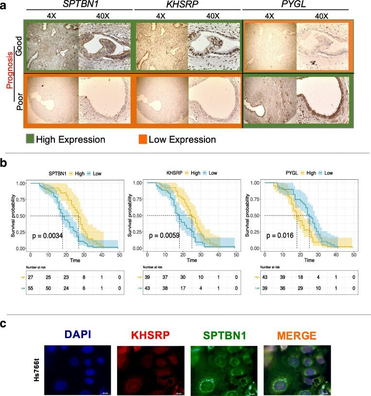
- Fig. 4 SPTBN1, KHSRP and PYGL as prognostic markers for resected pancreatic cancer patients. a . Immunohistochemistry validation of SPTBN1, KHSRP and PYGL on TMAs of 82 patients. b . Kaplan-Meier curves for SPTBN1, KHSRP and PYGL with p values 0.0034, 0.0059 and 0.016, respectively. c . Immunofluorescence of KHSRP (red) and SPTBN1 (green) in Hs766t cells