Explore
 Validate
 Learn
Learn Western blot
 Immunoprecipitation
Antibody data
- Antibody Data
- Antigen structure
- References [0]
- Comments [0]
- Validations
- Western blot [1]
- Immunocytochemistry [1]
Submit
Validation data
Reference
Comment
Report error
- Product number
- GTX15835 - Provider product page

- Provider
- GeneTex
- Product name
- Synaptojanin 1 antibody [AC1]
- Antibody type
- Monoclonal
- Reactivity
- Human, Rat
- Host
- Mouse
No comments: Submit comment
Supportive validation
- Submitted by
- GeneTex (provider)
- Main image
- Experimental details
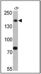
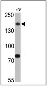
- Western blot analysis of Synaptojanin 1 in 25 ug of C6 cell lysates. Proteins were transferred to a PVDF membrane and blocked at 4¢XC overnight. The membrane was probed with Synaptojanin 1 antibody [AC1] at a dilution of 1:500 overnight at 4¢XC, washed in TBST, and probed with an HRP-conjugated secondary antibody. Chemiluminescent detection was performed .
Supportive validation
- Submitted by
- GeneTex (provider)
- Main image
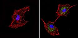
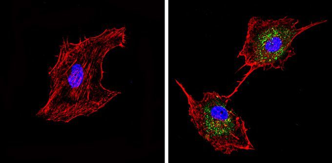
- Experimental details
- Immunofluorescent analysis of Synaptojanin 1 (green) in U-87 MG cells (right) compared to a negative control without primary antibody (left). Formalin-fixed cells were permeabilized with 0.1% Triton X-100 in TBS for 5-10 minutes and blocked with 3% BSA-PBS for 30 minutes at room temperature. Cells were probed with Synaptojanin 1 antibody [AC1] in 3% BSA-PBS at a dilution of 1:50 and incubated overnight at 4¢XC in a humidified chamber. Cells were washed with PBST and incubated with a proper secondary antibody. F-actin (red) was stained with a flourescent red phalloidin and nuclei (blue) were stained with Hoechst or DAPI. Images were taken at a magnification of 60x.