Explore
 Validate
 Learn
Learn Western blot
 ELISA
ELISA Immunohistochemistry
Antibody data
- Antibody Data
- Antigen structure
- References [0]
- Comments [0]
- Validations
- Western blot [2]
- Immunocytochemistry [2]
Submit
Validation data
Reference
Comment
Report error
- Product number
- PA5-112733 - Provider product page

- Provider
- Invitrogen Antibodies
- Product name
- Synaptojanin 1 Polyclonal Antibody
- Antibody type
- Polyclonal
- Antigen
- Recombinant full-length protein
- Reactivity
- Human
- Host
- Rabbit
- Isotype
- IgG
- Vial size
- 100 µg
- Concentration
- 4.6 mg/mL
- Storage
- -20°C or -80°C if preferred
No comments: Submit comment
Supportive validation
- Submitted by
- Invitrogen Antibodies (provider)
- Main image

- Experimental details
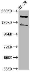
- Western blot analysis of Synaptojanin 1 using a polyclonal antibody (Product # PA5-112733) at a dilution of 2 µg/mL. A goat anti-rabbit polyclonal secondary antibody at a dilution of 1:50,000 was used for detection in HT29 whole cell lysate. Observed band: 174 kDa.
- Submitted by
- Invitrogen Antibodies (provider)
- Main image

- Experimental details
- Western blot analysis of Synaptojanin 1 using a polyclonal antibody (Product # PA5-112733) at a dilution of 2 µg/mL. A goat anti-rabbit polyclonal secondary antibody at a dilution of 1:50,000 was used for detection in HT29 whole cell lysate. Observed band: 174 kDa.

Supportive validation
- Submitted by
- Invitrogen Antibodies (provider)
- Main image
- Experimental details
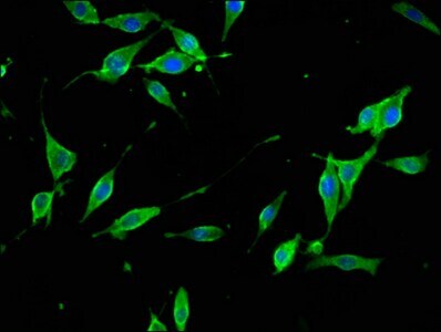
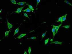
- Immunocytochemichal analysis of Synaptojanin 1 in NIH/3T3 cells using a Polyclonal antibody (Product # PA5-112733) at a dilution of 1:133. The cells were fixed in 4% formaldehyde, permeabilized using 0.2% Triton X-100 and blocked in 10% normal Goat Serum. The cells were then incubated with the antibody overnight at 4°C. The secondary antibody was Alexa Fluor 488-congugated Goat Anti-Rabbit IgG(H+L). Cells were also counter-stained with DAPI.

- Submitted by
- Invitrogen Antibodies (provider)
- Main image

- Experimental details
- Immunocytochemichal analysis of Synaptojanin 1 in NIH/3T3 cells using a Polyclonal antibody (Product # PA5-112733) at a dilution of 1:133. The cells were fixed in 4% formaldehyde, permeabilized using 0.2% Triton X-100 and blocked in 10% normal Goat Serum. The cells were then incubated with the antibody overnight at 4°C. The secondary antibody was Alexa Fluor 488-congugated Goat Anti-Rabbit IgG(H+L). Cells were also counter-stained with DAPI.