Explore
 Validate
 Learn
Learn Immunocytochemistry
Antibody data
- Antibody Data
- Antigen structure
- References [5]
- Comments [0]
- Validations
- Immunocytochemistry [1]
- Immunohistochemistry [1]
Submit
Validation data
Reference
Comment
Report error
- Product number
- HPA007863 - Provider product page

- Provider
- Atlas Antibodies
- Proper citation
- Atlas Antibodies Cat#HPA007863, RRID:AB_1080135
- Product name
- Anti-SV2A
- Antibody type
- Polyclonal
- Description
- Polyclonal Antibody against Human SV2A, Gene description: synaptic vesicle glycoprotein 2A, Alternative Gene Names: KIAA0736, SV2, Validated applications: IHC, ICC, Uniprot ID: Q7L0J3, Storage: Store at +4°C for short term storage. Long time storage is recommended at -20°C.
- Reactivity
- Human
- Host
- Rabbit
- Conjugate
- Unconjugated
- Isotype
- IgG
- Vial size
- 100 µl
- Concentration
- 0.1 mg/ml
- Storage
- Store at +4°C for short term storage. Long time storage is recommended at -20°C.
- Handling
- The antibody solution should be gently mixed before use.
Submitted references Differentiation of two human neuroblastoma cell lines alters SV2 expression patterns
Levetiracetam treatment ameliorates LRRK2 pathological mutant phenotype
Lipid-dependent deposition of alpha-synuclein and Tau on neuronal Secretogranin II-positive vesicular membranes with age
Amyloid Beta A4 Precursor Protein-binding Family B Member 1 (FE65) Interactomics Revealed Synaptic Vesicle Glycoprotein 2A (SV2A) and Sarcoplasmic/Endoplasmic Reticulum Calcium ATPase 2 (SERCA2) as New Binding Proteins in the Human Brain
Immunofluorescence and fluorescent-protein tagging show high correlation for protein localization in mammalian cells
Lekholm E, Ceder M, Forsberg E, Schiöth H, Fredriksson R
Cellular & Molecular Biology Letters 2021;26(1)
Cellular & Molecular Biology Letters 2021;26(1)
Levetiracetam treatment ameliorates LRRK2 pathological mutant phenotype
Rassu M, Biosa A, Galioto M, Fais M, Sini P, Greggio E, Piccoli G, Crosio C, Iaccarino C
Journal of Cellular and Molecular Medicine 2019;23(12):8505-8510
Journal of Cellular and Molecular Medicine 2019;23(12):8505-8510
Lipid-dependent deposition of alpha-synuclein and Tau on neuronal Secretogranin II-positive vesicular membranes with age
Brekk O, Moskites A, Isacson O, Hallett P
Scientific Reports 2018;8(1)
Scientific Reports 2018;8(1)
Amyloid Beta A4 Precursor Protein-binding Family B Member 1 (FE65) Interactomics Revealed Synaptic Vesicle Glycoprotein 2A (SV2A) and Sarcoplasmic/Endoplasmic Reticulum Calcium ATPase 2 (SERCA2) as New Binding Proteins in the Human Brain
Nensa F, Neumann M, Schrötter A, Przyborski A, Mastalski T, Susdalzew S, Looβe C, Helling S, El Magraoui F, Erdmann R, Meyer H, Uszkoreit J, Eisenacher M, Suh J, Guénette S, Röhner N, Kögel D, Theiss C, Marcus K, Müller T
Molecular & Cellular Proteomics 2014;13(2):475-488
Molecular & Cellular Proteomics 2014;13(2):475-488
Immunofluorescence and fluorescent-protein tagging show high correlation for protein localization in mammalian cells
Stadler C, Rexhepaj E, Singan V, Murphy R, Pepperkok R, Uhlén M, Simpson J, Lundberg E
Nature Methods 2013;10(4):315-323
Nature Methods 2013;10(4):315-323
No comments: Submit comment
Supportive validation
- Submitted by
- Atlas Antibodies (provider)
- Main image
- Experimental details
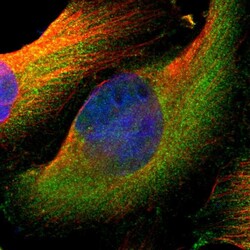
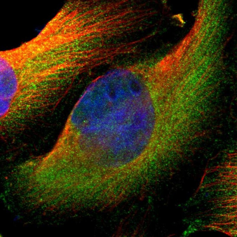
- Immunofluorescent staining of human cell line U-251 MG shows localization to cytosol.
- Sample type
- Human
Supportive validation
- Submitted by
- Atlas Antibodies (provider)
- Enhanced method
- Orthogonal validation
- Main image
- Experimental details
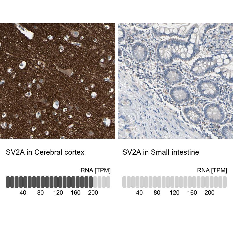
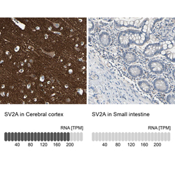
- Immunohistochemistry analysis in human cerebral cortex and small intestine tissues using HPA007863 antibody. Corresponding SV2A RNA-seq data are presented for the same tissues.
- Sample type
- Human
- Protocol
- Protocol