Explore
 Validate
 Learn
Learn Western blot
 Immunocytochemistry
 Immunoprecipitation
Antibody data
- Antibody Data
- Antigen structure
- References [1]
- Comments [0]
- Validations
- Immunocytochemistry [3]
- Immunohistochemistry [1]
Submit
Validation data
Reference
Comment
Report error
- Product number
- PA5-47563 - Provider product page

- Provider
- Invitrogen Antibodies
- Product name
- Brevican Polyclonal Antibody
- Antibody type
- Polyclonal
- Antigen
- Recombinant full-length protein
- Description
- In direct ELISAs, approximately 50% cross-reactivity with recombinant mouse Brevican is observed. Reconstitute at 0.2 mg/mL in sterile PBS.
- Reactivity
- Human, Rat
- Host
- Sheep
- Isotype
- IgG
- Vial size
- 100 μg
- Concentration
- 0.2 mg/mL
- Storage
- -20°C, Avoid Freeze/Thaw Cycles
Submitted references Increased matrix metalloproteinase levels and perineuronal net proteolysis in the HIV-infected brain; relevance to altered neuronal population dynamics.
Bozzelli PL, Caccavano A, Avdoshina V, Mocchetti I, Wu JY, Conant K
Experimental neurology 2020 Jan;323:113077
Experimental neurology 2020 Jan;323:113077
No comments: Submit comment
Supportive validation
- Submitted by
- Invitrogen Antibodies (provider)
- Main image

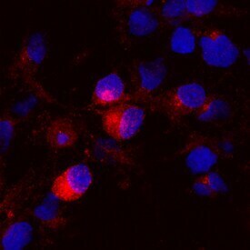
- Experimental details
- Immunocytochemistry analysis of Brevican in immersion fixed rat cortical stem cells differentiated for 7 days by growth factor withdrawal. Samples were incubated in Brevican polyclonal antibody (Product # PA5-47563) using a dilution of 10 µg/mL for 3 hours at room temperature followed by NorthernLights™ 557-conjugated Anti-Sheep IgG Secondary Antibody (red) and counterstained with DAPI (blue). Specific staining was localized to cytoplasm.

- Submitted by
- Invitrogen Antibodies (provider)
- Main image

- Experimental details
- Immunocytochemistry analysis of Brevican in immersion fixed rat cortical stem cells differentiated for 7 days by growth factor withdrawal. Samples were incubated in Brevican polyclonal antibody (Product # PA5-47563) using a dilution of 10 µg/mL for 3 hours at room temperature followed by NorthernLights™ 557-conjugated Anti-Sheep IgG Secondary Antibody (red) and counterstained with DAPI (blue). Specific staining was localized to cytoplasm.
Supportive validation
- Submitted by
- Invitrogen Antibodies (provider)
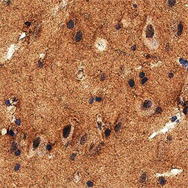
- Main image

- Experimental details
- Immunohistochemical analysis of Brevican in immersion fixed paraffin-embedded sections of human brain (cortex). Samples were incubated in Brevican polyclonal antibody (Product # PA5-47563) using a dilution of 5 µg/mL overnight at 4 °C. Tissue was stained using the Anti-Sheep HRP-DAB Cell & Tissue Staining Kit (brown) and counterstained with hematoxylin (blue). Specific staining was localized to neuropil.