 Explore
 Validate
 Learn
Learn Western blot
 Immunoprecipitation
Antibody data
- Antibody Data
- Antigen structure
- References [2]
- Comments [0]
- Validations
- Western blot [1]
- Immunocytochemistry [1]
- Immunohistochemistry [1]
Submit
Validation data
Reference
Comment
Report error
- Product number
- AF4009 - Provider product page

- Provider
- R&D Systems
- Product name
- Human/Rat Brevican Antibody
- Antibody type
- Polyclonal
- Description
- Antigen Affinity-purified. Detects human Brevican in direct ELISAs and Western blots. In direct ELISAs, less than 20% cross-reactivity with recombinant mouse Brevican is observed.
- Reactivity
- Human, Rat
- Host
- Sheep
- Conjugate
- Unconjugated
- Antigen sequence
AAH27971- Isotype
- IgG
- Vial size
- 100 ug
- Concentration
- LYOPH
- Storage
- Use a manual defrost freezer and avoid repeated freeze-thaw cycles. 12 months from date of receipt, -20 to -70 °C as supplied. 1 month, 2 to 8 °C under sterile conditions after reconstitution. 6 months, -20 to -70 °C under sterile conditions after reconstitution.
Submitted references Perineuronal Nets in Spinal Motoneurones: Chondroitin Sulphate Proteoglycan around Alpha Motoneurones.
Sensory experience-dependent formation of perineuronal nets and expression of Cat-315 immunoreactive components in the mouse somatosensory cortex.
Irvine SF, Kwok JCF
International journal of molecular sciences 2018 Apr 12;19(4)
International journal of molecular sciences 2018 Apr 12;19(4)
Sensory experience-dependent formation of perineuronal nets and expression of Cat-315 immunoreactive components in the mouse somatosensory cortex.
Ueno H, Suemitsu S, Okamoto M, Matsumoto Y, Ishihara T
Neuroscience 2017 Jul 4;355:161-174
Neuroscience 2017 Jul 4;355:161-174
No comments: Submit comment
Supportive validation
- Submitted by
- R&D Systems (provider)
- Main image
- Experimental details
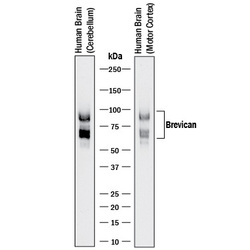
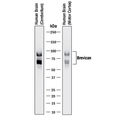
- Detection of Human Brevican by Western Blot. Western blot shows lysates of human brain (cerebellum) tissue and human brain (motor cortex) tissue. PVDF membrane was probed with 0.1 µg/mL of Sheep Anti-Human/Rat Brevican Antigen Affinity-purified Polyclonal Antibody (Catalog # AF4009) followed by HRP-conjugated Donkey Anti-Sheep IgG HRP-conjugated Antigen Affinity-purified Polyclonal Antibody (Catalog # HAF016) . Specific bands were detected for Brevican at approximately 60 and 90 kDa (as indicated). This experiment was conducted under reducing conditions and using Immunoblot Buffer Group 1.
Supportive validation
- Submitted by
- R&D Systems (provider)
- Main image

- Experimental details
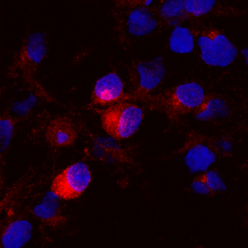
- Brevican in Rat Cortical Stem Cells. Brevican was detected in immersion fixed rat cortical stem cells differentiated for 7 days by growth factor withdrawal using Sheep Anti-Human/Rat Brevican Antigen Affinity-purified Polyclonal Antibody (Catalog # AF4009) at 10 µg/mL for 3 hours at room temperature. Cells were stained using the NorthernLights™ 557-conjugated Anti-Sheep IgG Secondary Antibody (red; Catalog # NL010) and counterstained with DAPI (blue). Specific staining was localized to cytoplasm. View our protocol for Fluorescent ICC Staining of Stem Cells on Coverslips.
Supportive validation
- Submitted by
- R&D Systems (provider)
- Main image
- Experimental details
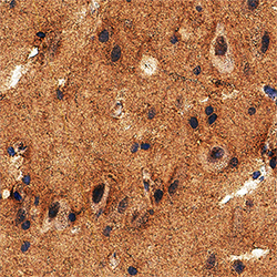
- Brevican in Human Brain. Brevican was detected in immersion fixed paraffin-embedded sections of human brain (cortex) using Sheep Anti-Human/Rat Brevican Antigen Affinity-purified Polyclonal Antibody (Catalog # AF4009) at 5 µg/mL overnight at 4 °C. Tissue was stained using the Anti-Sheep HRP-DAB Cell & Tissue Staining Kit (brown; Catalog # CTS019) and counterstained with hematoxylin (blue). Specific staining was localized to neuropil. View our protocol for Chromogenic IHC Staining of Paraffin-embedded Tissue Sections.
