Explore
 Validate
 Learn
Learn Western blot
 Immunohistochemistry
Antibody data
- Antibody Data
- Antigen structure
- References [2]
- Comments [0]
- Validations
- Western blot [1]
Submit
Validation data
Reference
Comment
Report error
- Product number
- PB9127 - Provider product page

- Provider
- Boster Biological Technology
- Product name
- Anti-Annexin A1/ANXA1 Antibody Picoband™
- Antibody type
- Polyclonal
- Description
- Polyclonal antibody for ANNEXIN I/ANXA1 detection. Host: Rabbit.Size: 100μg/vial. Tested applications: IHC-P. Reactive species: Human. ANNEXIN I/ANXA1 information: Molecular Weight: 38714 MW; Subcellular Localization: Nucleus . Cytoplasm . Cell projection, cilium . Basolateral cell membrane . Found in the cilium, nucleus and basolateral cell membrane of ciliated cells in the tracheal endothelium. Found in the cytoplasm of type II pneumocytes and alveolar macrophages.
- Reactivity
- Human, Mouse, Rat
- Host
- Rabbit
- Vial size
- 100μg/vial
- Concentration
- Add 0.2ml of distilled water will yield a concentration of 500ug/ml.
- Storage
- At -20°C for one year. After reconstitution, at 4°C for one month. It can also be aliquoted and stored frozen at -20°C for a longer time. Avoid repeated freezing and thawing.
- Handling
- Add 0.2ml of distilled water will yield a concentration of 500ug/ml.
Submitted references Annexin A1, A2, A4 and A5 play important roles in breast cancer, pancreatic cancer and laryngeal carcinoma, alone and/or synergistically.
Proteomic analysis of lanthanum citrate-induced apoptosis in human cervical carcinoma SiHa cells.
Deng S, Wang J, Hou L, Li J, Chen G, Jing B, Zhang X, Yang Z
Oncology letters 2013 Jan;5(1):107-112
Oncology letters 2013 Jan;5(1):107-112
Proteomic analysis of lanthanum citrate-induced apoptosis in human cervical carcinoma SiHa cells.
Shen L, Lan Z, Sun X, Shi L, Liu Q, Ni J
Biometals : an international journal on the role of metal ions in biology, biochemistry, and medicine 2010 Dec;23(6):1179-89
Biometals : an international journal on the role of metal ions in biology, biochemistry, and medicine 2010 Dec;23(6):1179-89
No comments: Submit comment
Supportive validation
- Submitted by
- Boster Biological Technology (provider)
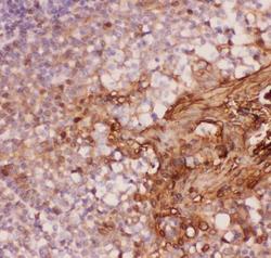
- Main image

- Experimental details
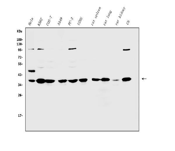
- Western blot analysis of Annexin A1/ANXA1 using anti-Annexin A1/ANXA1 antibody (PB9127). Electrophoresis was performed on a 5-20% SDS-PAGE gel at 70V (Stacking gel) / 90V (Resolving gel) for 2-3 hours. The sample well of each lane was loaded with 30ug of sample under reducing conditions. Lane 1: human Hela whole cell lysates, Lane 2: human K562 whole cell lysates, Lane 3: monkey COS-7 whole cell lysates, Lane 4: human A549 whole cell lysates, Lane 5: human PC-3 whole cell lysates, Lane 6: human U20S whole cell lysates, Lane 7: rat spleen tissue lysates, Lane 8: rat lung tissue lysates, Lane 9: rat kidney tissue lysates, Lane 10: rat C6 whole cell lysates. After Electrophoresis, proteins were transferred to a Nitrocellulose membrane at 150mA for 50-90 minutes. Blocked the membrane with 5% Non-fat Milk/ TBS for 1.5 hour at RT. The membrane was incubated with rabbit anti-Annexin A1/ANXA1 antigen affinity purified polyclonal antibody (Catalog # PB9127) at 0.5 μg/mL overnight at 4°C, then washed with TBS-0.1%Tween 3 times with 5 minutes each and probed with a goat anti-rabbit IgG-HRP secondary antibody at a dilution of 1:5000 for 1.5 hour at RT. The signal is developed using an Enhanced Chemiluminescent detection (ECL) kit (Catalog # EK1002) with Tanon 5200 system. A specific band was detected for Annexin A1/ANXA1 at approximately 39KD. The expected band size for Annexin A1/ANXA1 is at 39KD.
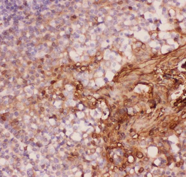
- Additional image