 Explore
 Validate
 Learn
Learn Western blot
 Immunocytochemistry
Antibody data
- Antibody Data
- Antigen structure
- References [2]
- Comments [0]
- Validations
- Western blot [1]
- Immunocytochemistry [1]
- Immunohistochemistry [1]
Submit
Validation data
Reference
Comment
Report error
- Product number
- AMAb90558 - Provider product page

- Provider
- Atlas Antibodies
- Proper citation
- Atlas Antibodies Cat#AMAb90558, RRID:AB_2665585
- Product name
- Anti-ANXA1
- Antibody type
- Monoclonal
- Description
- Monoclonal Antibody against Human ANXA1, Clone ID: CL0199, Gene description: annexin A1, Alternative Gene Names: ANX1, LPC1, Validated applications: ICC, IHC, WB, Uniprot ID: P04083, Storage: Store at +4°C for short term storage. Long time storage is recommended at -20°C.
- Reactivity
- Human
- Host
- Mouse
- Conjugate
- Unconjugated
- Isotype
- IgG
- Antibody clone number
- CL0199
- Vial size
- 100 µl
- Concentration
- 1.1 mg/ml
- Storage
- Store at +4°C for short term storage. Long time storage is recommended at -20°C.
- Handling
- The antibody solution should be gently mixed before use.
Submitted references Annexin A1 is a potential biomarker of bone metastasis in small cell lung cancer
Upregulation of annexin A1 protein expression in the intratumoral vasculature of human non–small-cell lung carcinoma and rodent tumor models
Chen P, Min J, Wu H, Zhang H, Wang C, Tan G, Zhang F
Oncology Letters 2020;21(2)
Oncology Letters 2020;21(2)
Upregulation of annexin A1 protein expression in the intratumoral vasculature of human non–small-cell lung carcinoma and rodent tumor models
Ulasov I, Allen K, Cann J, Zhao W, Peterson N, Lazzaro M, Zhong H, Wu H, Dall’Acqua W, Borrok M, Damschroder M, Tsui P, Li Q
PLOS ONE 2020;15(6):e0234268
PLOS ONE 2020;15(6):e0234268
No comments: Submit comment
Enhanced validation
- Submitted by
- Atlas Antibodies (provider)
- Enhanced method
- Genetic validation
- Main image
- Experimental details
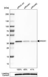
- Western blot analysis in U-251MG cells transfected with control siRNA, target specific siRNA probe #1 and #2, using Anti-ANXA1 antibody. Remaining relative intensity is presented. Loading control: Anti-PPIB.
- Sample type
- Human
- Protocol
- Protocol

Supportive validation
- Submitted by
- Atlas Antibodies (provider)
- Main image
- Experimental details
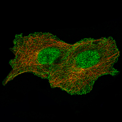
- Immunofluorescence staining of A549 cells using the Anti-ANXA1 monoclonal antibody, showing specific staining in the nucleus, cytosol and the plasma membrane in green. Microtubule- and nuclear probes are visualized in red and blue, respectively (where available).
- Sample type
- Human

Supportive validation
- Submitted by
- Atlas Antibodies (provider)
- Enhanced method
- Orthogonal validation
- Main image
- Experimental details
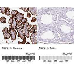
- Immunohistochemistry analysis in human placenta and testis tissues using AMAb90558 antibody. Corresponding ANXA1 RNA-seq data are presented for the same tissues.
- Sample type
- Human
- Protocol
- Protocol
