Explore
 Validate
 Learn
Learn Western blot
Antibody data
- Antibody Data
- Antigen structure
- References [2]
- Comments [0]
- Validations
- Western blot [2]
- Immunohistochemistry [1]
Submit
Validation data
Reference
Comment
Report error
- Product number
- AF3770 - Provider product page

- Provider
- R&D Systems
- Product name
- Human/Mouse/Rat Annexin A1 Antibody
- Antibody type
- Polyclonal
- Description
- Immunogen affinity purified. The antibody detects human, mouse and rat Annexin A1 in Western blots. In Western blots, goat anti-human/mouse/rat Annexin A1 does not cross-react with recombinant human Annexin A2, A3, A4, A6, A7, A8, A9, A10, A11 or A13.
- Reactivity
- Human, Mouse, Rat
- Host
- Goat
- Conjugate
- Unconjugated
- Antigen sequence
P04083- Isotype
- IgG
- Vial size
- 100 ug
- Concentration
- LYOPH
- Storage
- Use a manual defrost freezer and avoid repeated freeze-thaw cycles. 12 months from date of receipt, -20 to -70 °C as supplied. 1 month, 2 to 8 °C under sterile conditions after reconstitution. 6 months, -20 to -70 °C under sterile conditions after reconstitution.
Submitted references Phenotypic and Expressional Heterogeneity in the Invasive Glioma Cells.
A role for stroma-derived annexin A1 as mediator in the control of genetic susceptibility to T-cell lymphoblastic malignancies through prostaglandin E2 secretion.
Fayzullin A, Sandberg CJ, Spreadbury M, Saberniak BM, Grieg Z, Skaga E, Langmoen IA, Vik-Mo EO
Translational oncology 2019 Jan;12(1):122-133
Translational oncology 2019 Jan;12(1):122-133
A role for stroma-derived annexin A1 as mediator in the control of genetic susceptibility to T-cell lymphoblastic malignancies through prostaglandin E2 secretion.
Santos J, González-Sánchez L, Matabuena-Deyzaguirre M, Villa-Morales M, Cozar P, López-Nieva P, Fernández-Navarro P, Fresno M, Díaz-Muñoz MD, Guenet JL, Montagutelli X, Fernández-Piqueras J
Cancer research 2009 Mar 15;69(6):2577-87
Cancer research 2009 Mar 15;69(6):2577-87
No comments: Submit comment
Supportive validation
- Submitted by
- R&D Systems (provider)
- Main image
- Experimental details
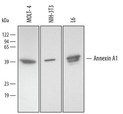
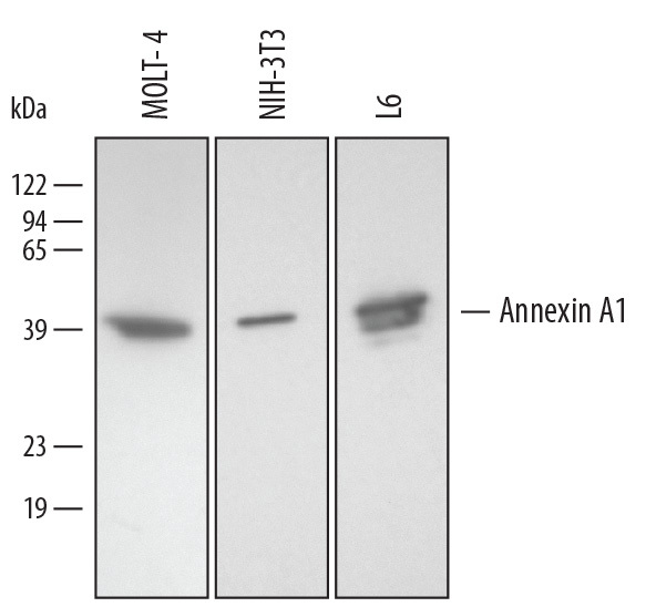
- Detection of Human/Mouse/Rat Annexin A1 by Western Blot. Western blot shows lysates of MOLT-4 human acute lymphoblastic leukemia cell line, NIH-3T3 mouse embryonic fibroblast cell line, and L6 rat myoblast cell line. PVDF membrane was probed with 0.2 µg/mL of Goat Anti-Human/Mouse/Rat Annexin A1 Antigen Affinity-purified Polyclonal Antibody (Catalog # AF3770) followed by HRP-conjugated Anti-Goat IgG Secondary Antibody (Catalog # HAF109). A specific band was detected for Annexin A1 at approximately 35-40 kDa (as indicated). This experiment was conducted under reducing conditions and using Immunoblot Buffer Group 2.
- Submitted by
- R&D Systems (provider)
- Main image
- Experimental details
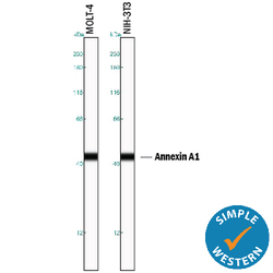
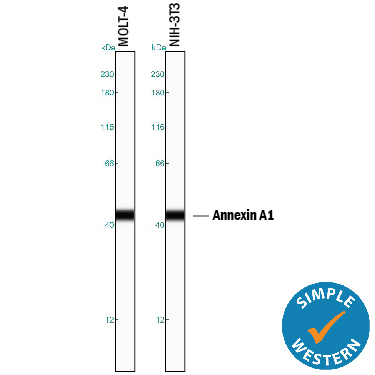
- Detection of Human and Mouse Annexin A1 by Simple WesternTM. Simple Western lane view shows lysates of MOLT-4 human acute lymphoblastic leukemia cell line and NIH-3T3 mouse embryonic fibroblast cell line, loaded at 0.2 mg/mL. A specific band was detected for Annexin A1 at approximately 44 kDa (as indicated) using 2 µg/mL of Goat Anti-Human/Mouse/Rat Annexin A1 Antigen Affinity-purified Polyclonal Antibody (Catalog # AF3770) followed by 1:50 dilution of HRP-conjugated Anti-Goat IgG Secondary Antibody (Catalog # HAF109). This experiment was conducted under reducing conditions and using the 12-230 kDa separation system.
Supportive validation
- Submitted by
- R&D Systems (provider)
- Main image
- Experimental details
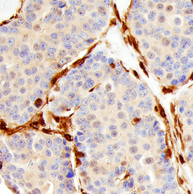
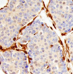
- Annexin A1/Annexin I in Human Breast. Annexin A1/Annexin I was detected in immersion fixed paraffin-embedded sections of human breast using Goat Anti-Human/Mouse/Rat Annexin A1/Annexin I Antigen Affinity-purified Polyclonal Antibody (Catalog # AF3770) at 10 µg/mL overnight at 4 °C. Before incubation with the primary antibody, tissue was subjected to heat-induced epitope retrieval using Antigen Retrieval Reagent-Basic (Catalog # CTS013). Tissue was stained using the Anti-Goat HRP-DAB Cell & Tissue Staining Kit (brown; Catalog # CTS008) and counter-stained with hematoxylin (blue). Specific staining was localized to stromal cells. View our protocol for Chromogenic IHC Staining of Paraffin-embedded Tissue Sections.