Explore
 Validate
 Learn
Learn Western blot
 Immunocytochemistry
Antibody data
- Antibody Data
- Antigen structure
- References [4]
- Comments [0]
- Validations
- Western blot [1]
- Immunocytochemistry [1]
- Immunohistochemistry [1]
Submit
Validation data
Reference
Comment
Report error
- Product number
- HPA011272 - Provider product page

- Provider
- Atlas Antibodies
- Proper citation
- Atlas Antibodies Cat#HPA011272, RRID:AB_1844884
- Product name
- Anti-ANXA1
- Antibody type
- Polyclonal
- Description
- Polyclonal Antibody against Human ANXA1, Gene description: annexin A1, Alternative Gene Names: ANX1, LPC1, Validated applications: ICC, IHC, WB, Uniprot ID: P04083, Storage: Store at +4°C for short term storage. Long time storage is recommended at -20°C.
- Reactivity
- Human, Mouse, Rat
- Host
- Rabbit
- Conjugate
- Unconjugated
- Isotype
- IgG
- Vial size
- 100 µl
- Concentration
- 0.1 mg/ml
- Storage
- Store at +4°C for short term storage. Long time storage is recommended at -20°C.
- Handling
- The antibody solution should be gently mixed before use.
Submitted references RNA-binding is an ancient trait of the Annexin family
Role of Annexin A1 in Squamous Cell Lung Cancer Progression
Nicotinamide Riboside Augments the Aged Human Skeletal Muscle NAD+ Metabolome and Induces Transcriptomic and Anti-inflammatory Signatures
Tumoral ANXA1 Is a Predictive Marker for Sunitinib Treatment of Renal Cancer Patients.
Patil S, Panchal V, Røstbø T, Romanyuk S, Hollås H, Brenk R, Grindheim A, Vedeler A
Frontiers in Cell and Developmental Biology 2023;11
Frontiers in Cell and Developmental Biology 2023;11
Role of Annexin A1 in Squamous Cell Lung Cancer Progression
Elakad O, Li Y, Gieser N, Yao S, Küffer S, Hinterthaner M, Danner B, von Hammerstein-Equord A, Ströbel P, Bohnenberger H, Mei J
Disease Markers 2021;2021
Disease Markers 2021;2021
Nicotinamide Riboside Augments the Aged Human Skeletal Muscle NAD+ Metabolome and Induces Transcriptomic and Anti-inflammatory Signatures
Elhassan Y, Kluckova K, Fletcher R, Schmidt M, Garten A, Doig C, Cartwright D, Oakey L, Burley C, Jenkinson N, Wilson M, Lucas S, Akerman I, Seabright A, Lai Y, Tennant D, Nightingale P, Wallis G, Manolopoulos K, Brenner C, Philp A, Lavery G
Cell Reports 2019;28(7):1717-1728.e6
Cell Reports 2019;28(7):1717-1728.e6
Tumoral ANXA1 Is a Predictive Marker for Sunitinib Treatment of Renal Cancer Patients.
Niinivirta M, Enblad G, Edqvist PH, Pontén F, Dragomir A, Ullenhag GJ
Journal of Cancer 2017;8(19):3975-3983
Journal of Cancer 2017;8(19):3975-3983
No comments: Submit comment
Enhanced validation
- Submitted by
- Atlas Antibodies (provider)
- Enhanced method
- Genetic validation
- Main image
- Experimental details
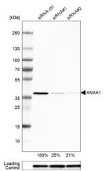
- Western blot analysis in U-251MG cells transfected with control siRNA, target specific siRNA probe #1 and #2, using Anti-ANXA1 antibody. Remaining relative intensity is presented. Loading control: Anti-PPIB.
- Sample type
- Human
- Protocol
- Protocol

Supportive validation
- Submitted by
- Atlas Antibodies (provider)
- Main image
- Experimental details
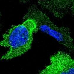
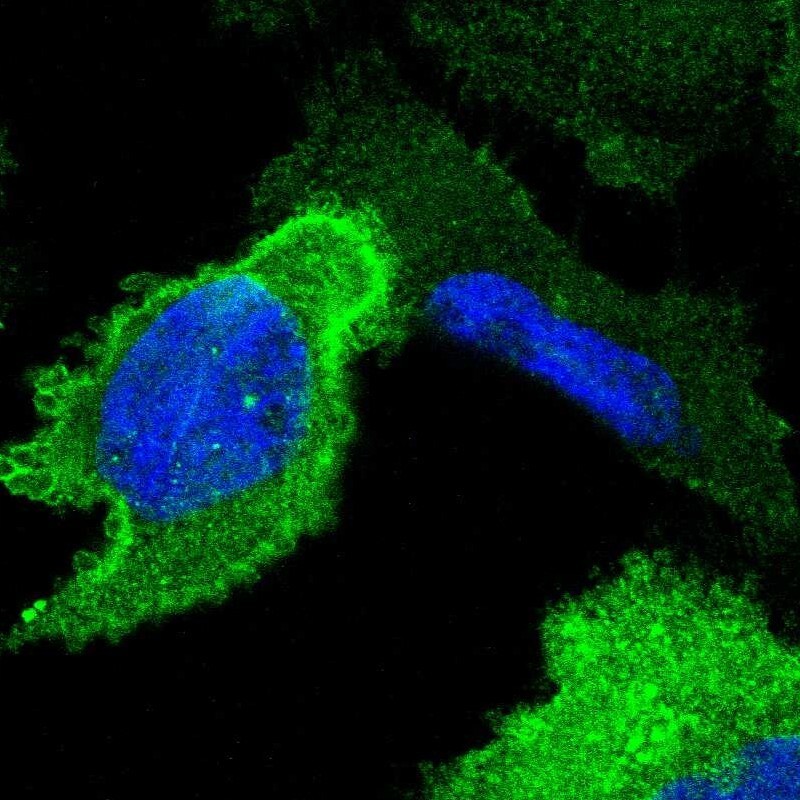
- Immunofluorescent staining of human cell line U-251 MG shows localization to plasma membrane & cytosol.
- Sample type
- Human
Supportive validation
- Submitted by
- Atlas Antibodies (provider)
- Enhanced method
- Orthogonal validation
- Main image

- Experimental details
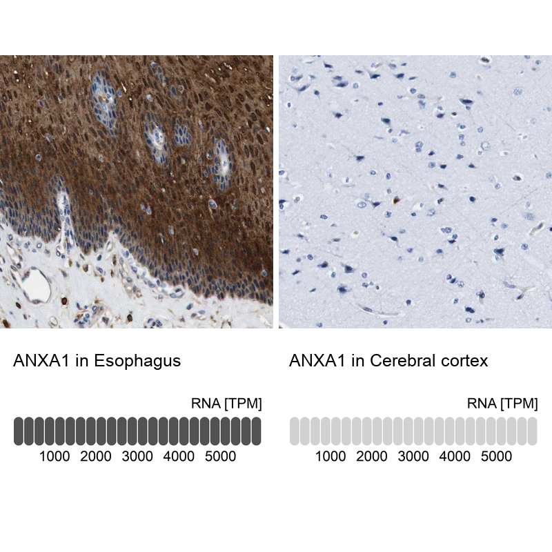
- Immunohistochemistry analysis in human esophagus and cerebral cortex tissues using HPA011272 antibody. Corresponding ANXA1 RNA-seq data are presented for the same tissues.
- Sample type
- Human
- Protocol
- Protocol