Explore
 Validate
 Learn
LearnPA1-742
antibody from Invitrogen Antibodies
Targeting: STXBP1
hUNC18, MUNC18-1, rbSec1, UNC18
 Western blot
 Immunocytochemistry
 Immunoprecipitation Immunohistochemistry Flow cytometry Other assay
Antibody data
- Antibody Data
- Antigen structure
- References [5]
- Comments [0]
- Validations
- Immunocytochemistry [3]
- Flow cytometry [1]
- Other assay [1]
Submit
Validation data
Reference
Comment
Report error
- Product number
- PA1-742 - Provider product page

- Provider
- Invitrogen Antibodies
- Product name
- MUNC18 Polyclonal Antibody
- Antibody type
- Polyclonal
- Antigen
- Synthetic peptide
- Description
- PA1-742 detects Munc-18 from human, rat and mouse tissues. PA1-742 has been successfully used in Western blot, immunohistochemistry, and immunoprecipitation procedures. By Western blot, this antibody detects an ~67 kDa protein representing Munc-18 from rat and mouse whole brain extract. PA1-742 immunizing peptide corresponds to amino acid residues 58-70 from human Munc-18. This sequence is completely conserved between human, mouse, rat, bovine, and canine Munc-18. PA1-742 immunizing peptide (Cat. # PEP-067) is available for use in neutralization and control experiments.
- Reactivity
- Human, Mouse, Rat
- Host
- Rabbit
- Isotype
- IgG
- Vial size
- 100 μg
- Concentration
- 1 mg/mL
- Storage
- -20°C, Avoid Freeze/Thaw Cycles
Submitted references Congenital Anophthalmia and Binocular Neonatal Enucleation Differently Affect the Proteome of Primary and Secondary Visual Cortices in Mice.
The Toll-like receptor-3 agonist polyinosinic:polycytidylic acid triggers nigrostriatal dopaminergic degeneration.
Apical targeting of syntaxin 3 is essential for epithelial cell polarity.
Munc18c regulates insulin-stimulated glut4 translocation to the transverse tubules in skeletal muscle.
A novel ubiquitous form of Munc-18 interacts with multiple syntaxins. Use of the yeast two-hybrid system to study interactions between proteins involved in membrane traffic.
Laramée ME, Smolders K, Hu TT, Bronchti G, Boire D, Arckens L
PloS one 2016;11(7):e0159320
PloS one 2016;11(7):e0159320
The Toll-like receptor-3 agonist polyinosinic:polycytidylic acid triggers nigrostriatal dopaminergic degeneration.
Deleidi M, Hallett PJ, Koprich JB, Chung CY, Isacson O
The Journal of neuroscience : the official journal of the Society for Neuroscience 2010 Dec 1;30(48):16091-101
The Journal of neuroscience : the official journal of the Society for Neuroscience 2010 Dec 1;30(48):16091-101
Apical targeting of syntaxin 3 is essential for epithelial cell polarity.
Sharma N, Low SH, Misra S, Pallavi B, Weimbs T
The Journal of cell biology 2006 Jun 19;173(6):937-48
The Journal of cell biology 2006 Jun 19;173(6):937-48
Munc18c regulates insulin-stimulated glut4 translocation to the transverse tubules in skeletal muscle.
Khan AH, Thurmond DC, Yang C, Ceresa BP, Sigmund CD, Pessin JE
The Journal of biological chemistry 2001 Feb 9;276(6):4063-9
The Journal of biological chemistry 2001 Feb 9;276(6):4063-9
A novel ubiquitous form of Munc-18 interacts with multiple syntaxins. Use of the yeast two-hybrid system to study interactions between proteins involved in membrane traffic.
Hata Y, Südhof TC
The Journal of biological chemistry 1995 Jun 2;270(22):13022-8
The Journal of biological chemistry 1995 Jun 2;270(22):13022-8
No comments: Submit comment
Supportive validation
- Submitted by
- Invitrogen Antibodies (provider)
- Main image

- Experimental details
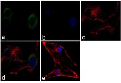
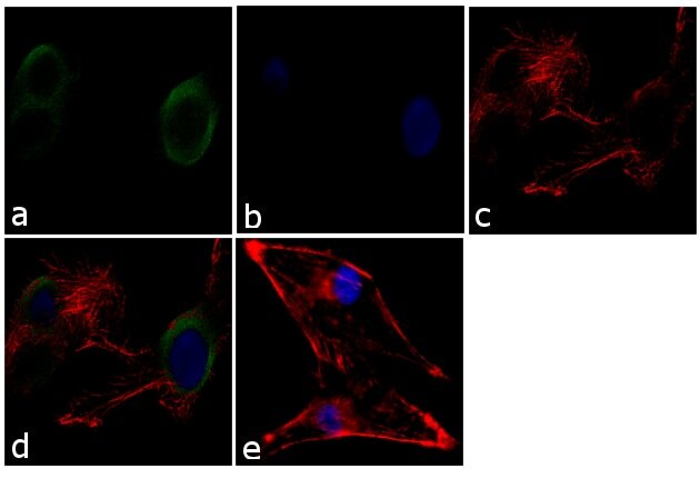
- Immunofluorescence analysis of MUNC18 was performed using 70% confluent log phase U-87 MG cells. The cells were fixed with 4% paraformaldehyde for 10 minutes, permeabilized with 0.1% Triton™ X-100 for 10 minutes, and blocked with 1% BSA for 1 hour at room temperature. The cells were labeled with MUNC18 Rabbit Polyclonal Antibody (Product # PA1-742) at 2µg/ml in 0.1% BSA and incubated for 3 hours at room temperature and then labeled with Goat anti-Rabbit IgG (H+L) Superclonal™ Secondary Antibody, Alexa Fluor® 488 conjugate (Product # A27034) at a dilution of 1:2000 for 45 minutes at room temperature (Panel a: green). Nuclei (Panel b: blue) were stained with SlowFade® Gold Antifade Mountant with DAPI (Product # S36938). F-actin (Panel c: red) was stained with Alexa Fluor® 555 Rhodamine Phalloidin (Product # R415, 1:300). Panel d represents the merged image showing cytoplasmic localization. Panel e shows the no primary antibody control. The images were captured at 60X magnification.

- Submitted by
- Invitrogen Antibodies (provider)
- Main image
- Experimental details
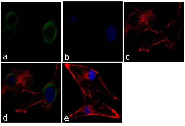
- Immunofluorescence analysis of MUNC18 was performed using 70% confluent log phase U-87 MG cells. The cells were fixed with 4% paraformaldehyde for 10 minutes, permeabilized with 0.1% Triton™ X-100 for 10 minutes, and blocked with 1% BSA for 1 hour at room temperature. The cells were labeled with MUNC18 Rabbit Polyclonal Antibody (Product # PA1-742) at 2µg/mL in 0.1% BSA and incubated for 3 hours at room temperature and then labeled with Goat anti-Rabbit IgG (H+L) Superclonal™ Secondary Antibody, Alexa Fluor® 488 conjugate (Product # A27034) at a dilution of 1:2000 for 45 minutes at room temperature (Panel a: green). Nuclei (Panel b: blue) were stained with SlowFade® Gold Antifade Mountant with DAPI (Product # S36938). F-actin (Panel c: red) was stained with Alexa Fluor® 555 Rhodamine Phalloidin (Product # R415, 1:300). Panel d represents the merged image showing cytoplasmic localization. Panel e shows the no primary antibody control. The images were captured at 60X magnification.
- Submitted by
- Invitrogen Antibodies (provider)
- Main image
- Experimental details
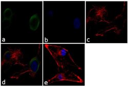
- Immunofluorescence analysis of MUNC18 was performed using 70% confluent log phase U-87 MG cells. The cells were fixed with 4% paraformaldehyde for 10 minutes, permeabilized with 0.1% Triton™ X-100 for 10 minutes, and blocked with 1% BSA for 1 hour at room temperature. The cells were labeled with MUNC18 Rabbit Polyclonal Antibody (Product # PA1-742) at 2µg/mL in 0.1% BSA and incubated for 3 hours at room temperature and then labeled with Goat anti-Rabbit IgG (Heavy Chain) Superclonal™ Secondary Antibody, Alexa Fluor® 488 conjugate (Product # A27034) at a dilution of 1:2000 for 45 minutes at room temperature (Panel a: green). Nuclei (Panel b: blue) were stained with SlowFade® Gold Antifade Mountant with DAPI (Product # S36938). F-actin (Panel c: red) was stained with Alexa Fluor® 555 Rhodamine Phalloidin (Product # R415, 1:300). Panel d represents the merged image showing cytoplasmic localization. Panel e shows the no primary antibody control. The images were captured at 60X magnification.
Supportive validation
- Submitted by
- Invitrogen Antibodies (provider)
- Main image
- Experimental details
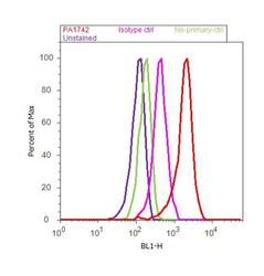
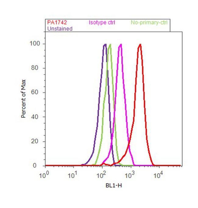
- Flow cytometry analysis of MUNC18 was done on U-87 MG cells. Cells were fixed with 70% ethanol for 10 minutes, permeabilized with 0.25% Triton™ X-100 for 20 minutes, and blocked with 5% BSA for 30 minutes at room temperature. Cells were labeled with MUNC18 Rabbit Polyclonal Antibody (PA1-742, red histogram) or with rabbit isotype control (pink histogram) at 3-5 µg/million cells in 2.5% BSA. After incubation at room temperature for 2 hours, the cells were labeled with Alexa Fluor® 488 Goat Anti-Rabbit Secondary Antibody (A11008) at a dilution of 1:400 for 30 minutes at room temperature. The representative 10,000 cells were acquired and analyzed for each sample using an Attune® Acoustic Focusing Cytometer. The purple histogram represents unstained control cells and the green histogram represents no-primary-antibody control..
Supportive validation
- Submitted by
- Invitrogen Antibodies (provider)
- Main image
- Experimental details
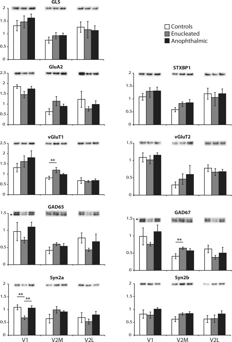
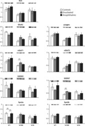
- Fig 2 Proteins involved in synaptic transmission. The expression of glutamine synthetase (GLS), glutamate AMPA 2 (GluA2), syntaxin binding protein 1 (STXBP1), glutamate vesicular transporter 1 (vGluT2) and 2 (vGluT2), glutamate decarboxylase 65 kb (GAD65) and 67 kb (GAD67), synapsin 2a (Syn2a) and 2b (Syn2b) were analyzed by Western blotting. White bars are controls, grey bars are enucleated and black bars are anophthalmic mice. Results from V1 (left), V2M (middle) and V2L (right) are shown for each protein. ** P < 0.01.