 Explore
 Validate
 Learn
Learn Western blot
Antibody data
- Antibody Data
- Antigen structure
- References [0]
- Comments [0]
- Validations
- Western blot [4]
- Immunocytochemistry [2]
Submit
Validation data
Reference
Comment
Report error
- Product number
- PA5-30184 - Provider product page

- Provider
- Invitrogen Antibodies
- Product name
- MUNC18 Polyclonal Antibody
- Antibody type
- Polyclonal
- Antigen
- Recombinant protein fragment
- Description
- Recommended positive controls: 293T, A431, mouse brain, rat brain. Predicted reactivity: Mouse (100%), Rat (100%), Xenopus laevis (93%), Chicken (98%), Rhesus Monkey (100%), Bovine (100%). Store product as a concentrated solution. Centrifuge briefly prior to opening the vial.
- Reactivity
- Human, Mouse, Rat
- Host
- Rabbit
- Isotype
- IgG
- Vial size
- 100 µL
- Concentration
- 0.96 mg/mL
- Storage
- Store at 4°C short term. For long term storage, store at -20°C, avoiding freeze/thaw cycles.
No comments: Submit comment
Supportive validation
- Submitted by
- Invitrogen Antibodies (provider)
- Main image

- Experimental details
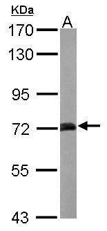
- Western blot analysis of STXBP1 using 20 µg of mouse brain lysate. Samples were loaded onto a 7.5% SDS-PAGE gel and probed with a STXBP1 polyclonal antibody (Product # PA5-30184) at a dilution of 1:10,000.
- Submitted by
- Invitrogen Antibodies (provider)
- Main image
- Experimental details
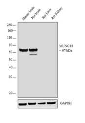
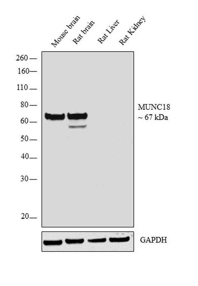
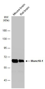
- Western blot analysis was performed on whole cell extract (30 µg lysate) of Mouse brain (Lane 1), Rat brain (Lane 2), Rat Liver (Lane 3), and Rat Kidney (Lane 4). The blot was probed with Anti-MUNC18 Polyclonal Antibody (Product # PA5-30184, 1:5000 dilution) and detected by chemiluminescence using Goat anti-Rabbit IgG (H+L) Superclonal™ Secondary Antibody, HRP conjugate (Product # A27036, 0.25 µg/mL, 1:4000 dilution). A 67 kDa band corresponding to MUNC18 was observed in the Mouse and Rat brain, while it was not detected in Rat Liver and Kidney which are reported negative for MUNC18 expression.
- Submitted by
- Invitrogen Antibodies (provider)
- Main image
- Experimental details
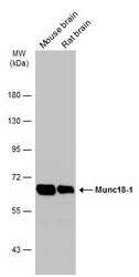
- Western Blot analysis of MUNC18 was performed by separating 50 µg of Various tissue extracts by 7.5% SDS-PAGE. Proteins were transferred to a membrane and probed with a MUNC18 Polyclonal Antibody (Product # PA5-30184) at a dilution of 1:10000. The HRP-conjugated anti-rabbit IgG antibody was used to detect the primary antibody.
- Submitted by
- Invitrogen Antibodies (provider)
- Main image
- Experimental details
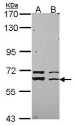
- Western Blot using MUNC18 Polyclonal Antibody (Product # PA5-30184). Sample (30 µg of whole cell lysate). Lane A: 293T. Lane B: A431 . 7.5% SDS PAGE. MUNC18 Polyclonal Antibody (Product # PA5-30184) diluted at 1:3,000.

Supportive validation
- Submitted by
- Invitrogen Antibodies (provider)
- Main image

- Experimental details
- Immunofluorescent analysis of STXBP1 in methanol-fixed A431 cells using a STXBP1 polyclonal antibody (Product # PA5-30184) (Green) at a 1:500 dilution. Alpha-tubulin filaments were labeled with Product # PA5-29281 (Red) at a 1:2000.
- Submitted by
- Invitrogen Antibodies (provider)
- Main image

- Experimental details
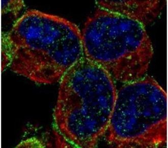
- Immunocytochemistry-Immunofluorescence analysis of MUNC18 was performed in DIV9 rat E18 primary cortical neurons fixed in 4% paraformaldehyde at RT for 15 min. Green: MUNC18 Polyclonal Antibody (Product # PA5-30184) diluted at 1:500. Red: beta Tubulin 3/ Tuj1, stained by beta Tubulin 3/ Tuj1 antibody. Blue: Fluoroshield with DAPI.
