Explore
 Validate
 Learn
Learn Western blot
Antibody data
- Antibody Data
- Antigen structure
- References [3]
- Comments [0]
- Validations
- Western blot [7]
- Immunocytochemistry [1]
Submit
Validation data
Reference
Comment
Report error
- Product number
- GTX111039 - Provider product page

- Provider
- GeneTex
- Proper citation
- GeneTex Cat#GTX111039, RRID:AB_10721697
- Product name
- Calcineurin A antibody
- Antibody type
- Polyclonal
- Reactivity
- Human, Rat
- Host
- Rabbit
Submitted references Effects of anoxic exposure on the nuclear factor of activated T cell (NFAT) transcription factors in the stress-tolerant wood frog.
Regulation of gene expression by NFAT transcription factors in hibernating ground squirrels is dependent on the cellular environment.
Elevated activation of CaMKIIα in the CPEB3-knockout hippocampus impairs a specific form of NMDAR-dependent synaptic depotentiation.
Al-Attar R, Storey KB
Cell biochemistry and function 2018 Dec;36(8):420-430
Cell biochemistry and function 2018 Dec;36(8):420-430
Regulation of gene expression by NFAT transcription factors in hibernating ground squirrels is dependent on the cellular environment.
Zhang Y, Storey KB
Cell stress & chaperones 2016 Sep;21(5):883-94
Cell stress & chaperones 2016 Sep;21(5):883-94
Elevated activation of CaMKIIα in the CPEB3-knockout hippocampus impairs a specific form of NMDAR-dependent synaptic depotentiation.
Huang WH, Chao HW, Tsai LY, Chung MH, Huang YS
Frontiers in cellular neuroscience 2014;8:367
Frontiers in cellular neuroscience 2014;8:367
No comments: Submit comment
Supportive validation
- Submitted by
- GeneTex (provider)
- Main image
- Experimental details
- Sample (30 ug of whole cell lysate) A: IMR32 7.5% SDS PAGE GTX111039 diluted at 1:3000
- Validation comment
- WB

- Submitted by
- GeneTex (provider)
- Main image
- Experimental details
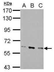
- Sample (30 ug of whole cell lysate) A: NIH-3T3 B: JC C: BCL-1 7.5% SDS PAGE GTX111039 diluted at 1:5000
- Validation comment
- WB
- Submitted by
- GeneTex (provider)
- Main image
- Experimental details
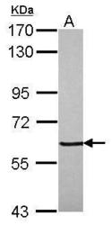
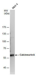
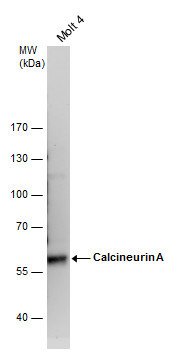
- Calcineurin A antibody detects Calcineurin A protein by western blot analysis. Whole cell extracts (30 £gg) was separated by 7.5 % SDS-PAGE, and blotted with Calcineurin A antibody (GTX111039) diluted by 1:3000
- Submitted by
- GeneTex (provider)
- Main image
- Experimental details
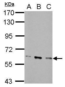
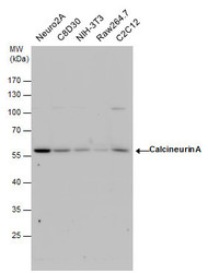
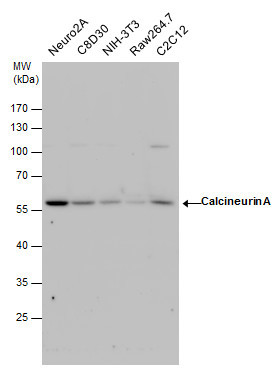
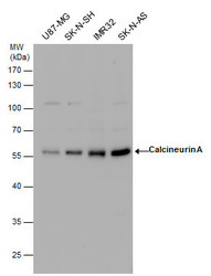
- Calcineurin A antibody detects Calcineurin A protein by western blot analysis. Various whole cell extracts (30 £gg) were separated by 10% SDS-PAGE, and the membrane was blotted with Calcineurin A antibody (GTX111039) diluted by 1:3000.
- Submitted by
- GeneTex (provider)
- Main image
- Experimental details
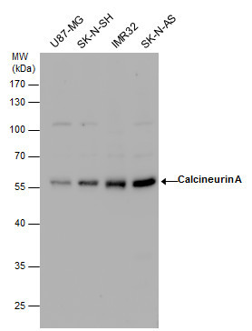
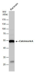
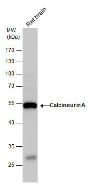
- Calcineurin A antibody detects Calcineurin A protein by western blot analysis. Rat tissue extracts (50 £gg) was separated by 10 % SDS-PAGE, and the membrane was blotted with Calcineurin A antibody (GTX111039) diluted by 1:3000.
- Submitted by
- GeneTex (provider)
- Main image
- Experimental details
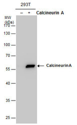
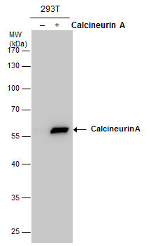
- Calcineurin A antibody detects Calcineurin A protein by western blot analysis. Non-transfected (-) and Calcineurin A-transfected (+, ) 293T whole cell extracts (30 £gg) were separated by 10% SDS-PAGE, and the membrane was blotted with Calcineurin A antibody (GTX111039) at a dilution of 1:5000.
Supportive validation
- Submitted by
- GeneTex (provider)
- Main image
- Experimental details
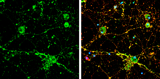
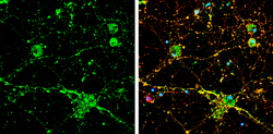
- Calcineurin A antibody detects Calcineurin A protein by immunofluorescent analysis.Sample: DIV9 rat E18 primary cortical neuron cells were fixed in 4% paraformaldehyde at RT for 15 min.Green: Calcineurin A stained by Calcineurin A antibody (GTX111039) diluted at 1:500.Red: beta Tubulin 3/ Tuj1, stained by beta Tubulin 3/ Tuj1 antibody [GT1338] (GTX631831) diluted at 1:500.Blue: Fluoroshield with DAPI (GTX30920).