Explore
 Validate
 Learn
Learn Western blot
Antibody data
- Antibody Data
- Antigen structure
- References [1]
- Comments [0]
- Validations
- Western blot [5]
- Immunocytochemistry [2]
- Immunohistochemistry [1]
Submit
Validation data
Reference
Comment
Report error
- Product number
- GTX111207 - Provider product page

- Provider
- GeneTex
- Proper citation
- GeneTex Cat#GTX111207, RRID:AB_1951358
- Product name
- Calcineurin A antibody
- Antibody type
- Polyclonal
- Reactivity
- Human, Mouse, Rat
- Host
- Rabbit
Submitted references Proteomic analysis of proteins responsible for the development of doxorubicin resistance in human uterine cancer cells.
Lin ST, Chou HC, Chang SJ, Chen YW, Lyu PC, Wang WC, Chang MD, Chan HL
Journal of proteomics 2012 Oct 22;75(18):5822-47
Journal of proteomics 2012 Oct 22;75(18):5822-47
No comments: Submit comment
Supportive validation
- Submitted by
- GeneTex (provider)
- Main image

- Experimental details
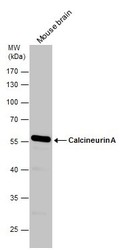
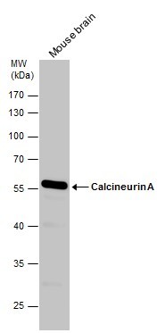
- Sample (50 ug of whole cell lysate) A: Mouse brain 7.5% SDS PAGE GTX111207 diluted at 1:1000
- Validation comment
- WB
- Submitted by
- GeneTex (provider)
- Main image
- Experimental details
- Sample (30 ug of whole cell lysate) A: Molt-4 (GTX27912) 7.5% SDS PAGE GTX111207 diluted at 1:1000
- Validation comment
- WB

- Submitted by
- GeneTex (provider)
- Main image
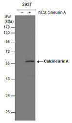
- Experimental details
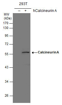
- Calcineurin A antibody detects Calcineurin A protein by western blot analysis. Non-transfected (-) and Calcineurin A-transfected (+) 293T whole cell extracts (30 ?g) were separated by 10% SDS-PAGE, and the membrane was blotted with Calcineurin A antibody (GTX111207) diluted at 1:1000. The HRP-conjugated anti-rabbit IgG antibody (GTX213110-01) was used to detect the primary antibody.
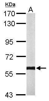
- Submitted by
- GeneTex (provider)
- Main image
- Experimental details
- Mouse tissue extract (50 ?g) was separated by 10% SDS-PAGE, and the membrane was blotted with Calcineurin A antibody (GTX111207) diluted at 1:500. The HRP-conjugated anti-rabbit IgG antibody (GTX213110-01) was used to detect the primary antibody.
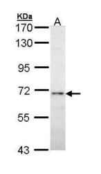
- Submitted by
- GeneTex (provider)
- Main image
- Experimental details
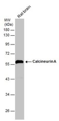
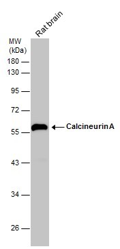
- Rat tissue extract (50 ?g) was separated by 10% SDS-PAGE, and the membrane was blotted with Calcineurin A antibody (GTX111207) diluted at 1:500. The HRP-conjugated anti-rabbit IgG antibody (GTX213110-01) was used to detect the primary antibody.
Supportive validation
- Submitted by
- GeneTex (provider)
- Main image
- Experimental details
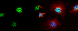
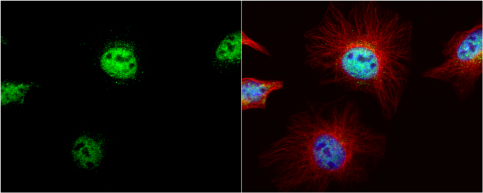
- Calcineurin A antibody detects Calcineurin A protein at nucleus by immunofluorescent analysis.Sample: HeLa cells were fixed in 4% paraformaldehyde at RT for 15 min.Green: Calcineurin A protein stained by Calcineurin A antibody (GTX111207) diluted at 1:200.Red: alpha Tubulin, a cytoskeleton marker, stained by alpha Tubulin antibody [B-5-1-2] (GTX11304) diluted at 1:10000.Blue: Hoechst 33342 staining.
- Submitted by
- GeneTex (provider)
- Main image
- Experimental details
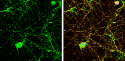
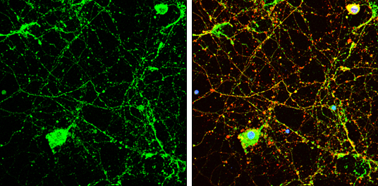
- Calcineurin A antibody detects Calcineurin A protein by immunofluorescent analysis.Sample: DIV9 rat E18 primary cortical neuron cells were fixed in 4% paraformaldehyde at RT for 15 min.Green: Calcineurin A stained by Calcineurin A antibody (GTX111207) diluted at 1:500.Red: beta Tubulin 3/ Tuj1, stained by beta Tubulin 3/ Tuj1 antibody [GT1338] (GTX631831) diluted at 1:500.Blue: Fluoroshield with DAPI (GTX30920).
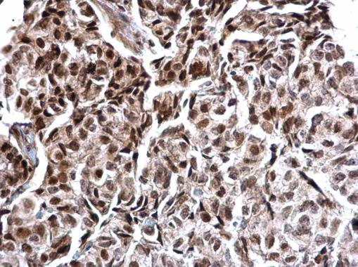
Supportive validation
- Submitted by
- GeneTex (provider)
- Main image
- Experimental details
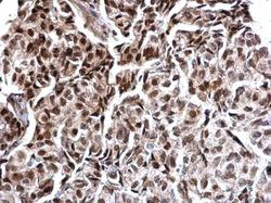
- Calcineurin A antibody detects Calcineurin A protein at nucleus on human breast carcinoma by immunohistochemical analysis. Sample: Paraffin-embedded human breast carcinoma. Calcineurin A antibody (GTX111207) dilution: 1:500.