 Explore
 Validate
 Learn
Learn Western blot
 Immunoprecipitation
Antibody data
- Antibody Data
- Antigen structure
- References [0]
- Comments [0]
- Validations
- Western blot [2]
- Immunocytochemistry [2]
- Immunohistochemistry [1]
Submit
Validation data
Reference
Comment
Report error
- Product number
- PA5-77819 - Provider product page

- Provider
- Invitrogen Antibodies
- Product name
- Calcineurin A Polyclonal Antibody
- Antibody type
- Polyclonal
- Antigen
- Synthetic peptide
- Reactivity
- Human, Mouse, Rat, Canine, Rabbit
- Host
- Rabbit
- Isotype
- IgG
- Vial size
- 100 µg
- Concentration
- 1 mg/mL
- Storage
- -20°C
No comments: Submit comment
Supportive validation
- Submitted by
- Invitrogen Antibodies (provider)
- Main image
- Experimental details
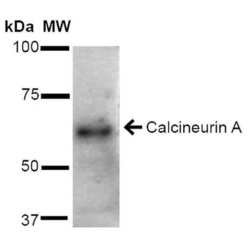
- Western blot analysis of Calcineurin A in rat brain cell lysates with 15 µg of sample. The sample was incubated with Calcineurin A polyclonal antibody (Product # PA5-77819) using a dilution of 1:1000 (2 hours at 4°C), and followed by Goat-Anti-Rabbit HRP (1 hour at RT) at a dilution of 1:2000 and ECL development (5 min at RT). Samples were arranged as follows: Lane 1: Molecular Weight Ladder (MW), Lane 2: Rat Brain cell lysates.
- Submitted by
- Invitrogen Antibodies (provider)
- Main image

- Experimental details
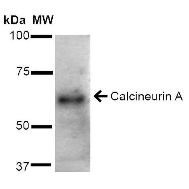
- Western blot was performed using Anti-Calcineurin A Rabbit Polyclonal Antibody (Product # PA5-77819) and 58 kDa band corresponding to Calcineurin A was observed across tissues tested except mouse thymus and mouse heart which are reported to be negative. An uncharacterized band at ~32 kDa was observed in positive tissues. Whole cell extracts (30 µg lysate) of Mouse Cerebellum (Lane 1), Mouse Brain (Lane 2), Rat Brain (Lane 3), Mouse Thymus (Lane 4) and Mouse Heart (Lane 5) were electrophoresed using NuPAGE™ 4-12% Bis-Tris Protein Gel (Product # NP0322BOX). Resolved proteins were then transferred onto a nitrocellulose membrane (Product # IB23001) by iBlot® 2 Dry Blotting System (Product # IB21001). The blot was probed with the primary antibody (1:1000 dilution) and detected by chemiluminescence with Goat anti-Rabbit IgG (H+L) Superclonal™ Recombinant Secondary Antibody, HRP (Product # A27036, 1:4000 dilution) using the iBright FL 1000 (Product # A32752). Chemiluminescent detection was performed using Novex® ECL Chemiluminescent Substrate Reagent Kit (Product # WP20005).

Supportive validation
- Submitted by
- Invitrogen Antibodies (provider)
- Main image

- Experimental details
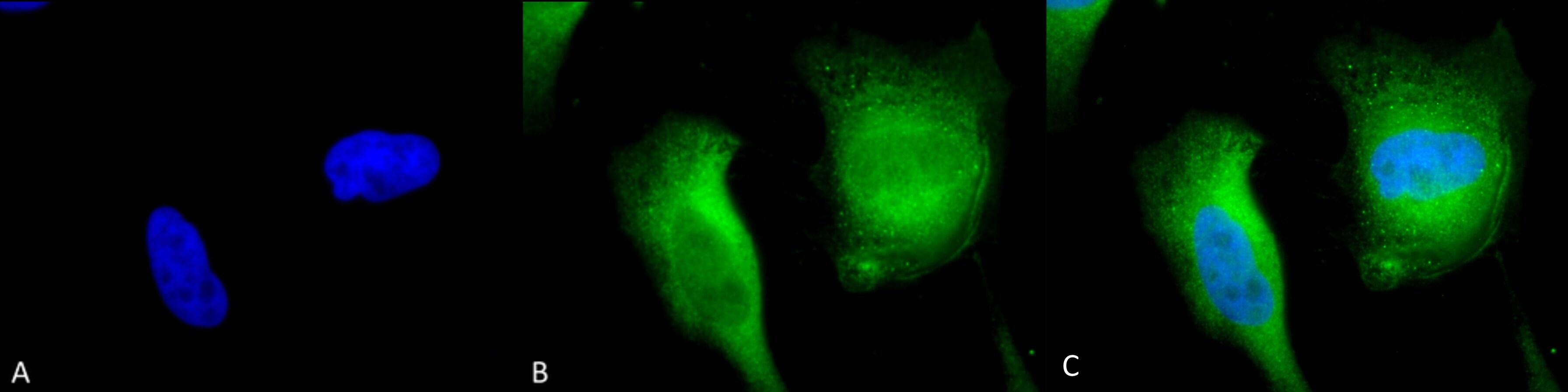
- Immunofluorescent analysis of Calcineurin A in HeLa cells. Sample was fixed using 2% formaldehyde (20 min at RT), incubated with Calcineurin A polyclonal antibody (Product # PA5-77819) using a dilution of 1:120 (12 hours at 4°C), and followed by APC Goat Anti-Rabbit (red) at 1:200 for 2 hours at RT. Counterstain: DAPI (blue) nuclear stain at 1:40000 for 2 hours at RT. Localization: Cell membrane. Cytoplasm. Magnification: 20x. Images are as follows: (A) DAPI (blue) nuclear stain. (B) Anti-Calcineurin A Antibody. (C) Composite.

- Submitted by
- Invitrogen Antibodies (provider)
- Main image

- Experimental details
- Immunofluorescent analysis of Calcineurin A in human HeLa cells. Sample was incubated with Calcineurin A polyclonal antibody (Product # PA5-77819) using a dilution of 1:100 (1 hour at RT), and followed by FITC Goat Anti-Mouse (green) secondary antibody at a dilution of 1:200, 1:1000 and 1:5000.
Supportive validation
- Submitted by
- Invitrogen Antibodies (provider)
- Main image

- Experimental details
- Immunohistochemistry analysis of Calcineurin A in mouse back skin. Sample was fixed using Bouin's fixative solution, incubated with Calcineurin A polyclonal antibody (Product # PA5-77819) using a dilution of 1:100 (1 hour at RT), and followed by FITC Goat Anti-Mouse (green) (1 hour at RT) at a dilution of 1:50.
