 Explore
 Validate
 Learn
Learn Western blot
 Immunocytochemistry
Antibody data
- Antibody Data
- Antigen structure
- References [1]
- Comments [0]
- Validations
- Immunocytochemistry [2]
- Immunohistochemistry [1]
Submit
Validation data
Reference
Comment
Report error
- Product number
- PA5-29255 - Provider product page

- Provider
- Invitrogen Antibodies
- Product name
- Calcineurin A Polyclonal Antibody
- Antibody type
- Polyclonal
- Antigen
- Recombinant full-length protein
- Description
- Recommended positive controls: human PPP3CA-transfected 293T, mouse brain, Rat brain. Predicted reactivity: Mouse (100%), Rat (100%), Zebrafish (93%), Xenopus laevis (98%), Dog (100%), Pig (99%), Rabbit (100%), Rhesus Monkey (100%), Bovine (100%). Store product as a concentrated solution. Centrifuge briefly prior to opening the vial.
- Reactivity
- Human, Mouse, Rat
- Host
- Rabbit
- Isotype
- IgG
- Vial size
- 100 μL
- Concentration
- 0.77 mg/mL
- Storage
- Store at 4°C short term. For long term storage, store at -20°C, avoiding freeze/thaw cycles.
Submitted references AKT inhibition-mediated dephosphorylation of TFE3 promotes overactive autophagy independent of MTORC1 in cadmium-exposed bone mesenchymal stem cells.
Pi H, Li M, Zou L, Yang M, Deng P, Fan T, Liu M, Tian L, Tu M, Xie J, Chen M, Li H, Xi Y, Zhang L, He M, Lu Y, Chen C, Zhang T, Wang Z, Yu Z, Gao F, Zhou Z
Autophagy 2019 Apr;15(4):565-582
Autophagy 2019 Apr;15(4):565-582
No comments: Submit comment
Supportive validation
- Submitted by
- Invitrogen Antibodies (provider)
- Main image
- Experimental details
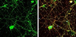
- Immunocytochemistry-Immunofluorescence analysis of Calcineurin A was performed in DIV9 rat E18 primary cortical neuron cells fixed in 4% paraformaldehyde at RT for 15 min. Green: Calcineurin A Polyclonal Antibody (Product # PA5 29255) diluted at 1:500. Red: beta Tubulin 3/ Tuj1, stained by beta Tubulin 3/ Tuj1 antibody. Blue: Fluoroshield with DAPI.
- Submitted by
- Invitrogen Antibodies (provider)
- Main image

- Experimental details
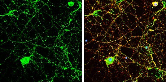
- Immunocytochemistry-Immunofluorescence analysis of Calcineurin A was performed in DIV9 rat E18 primary cortical neuron cells fixed in 4% paraformaldehyde at RT for 15 min. Green: Calcineurin A Polyclonal Antibody (Product # PA5 29255) diluted at 1:500. Red: beta Tubulin 3/ Tuj1, stained by beta Tubulin 3/ Tuj1 antibody. Blue: Fluoroshield with DAPI.

Supportive validation
- Submitted by
- Invitrogen Antibodies (provider)
- Main image
- Experimental details
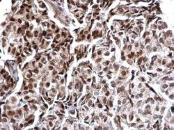
- Calcineurin A Polyclonal Antibody detects Calcineurin A protein at nucleus on human breast carcinoma by immunohistochemical analysis. Sample: Paraffin-embedded human breast carcinoma. Calcineurin A Polyclonal Antibody (Product # PA5-29255) dilution: 1:500. Antigen Retrieval: EDTA based buffer, pH 8.0, 15 min.
