Antibody data
- Antibody Data
- Antigen structure
- References [1]
- Comments [0]
- Validations
- Western blot [1]
- Immunohistochemistry [1]
Submit
Validation data
Reference
Comment
Report error
- Product number
- PA5-47468 - Provider product page

- Provider
- Invitrogen Antibodies
- Product name
- Neurofascin Polyclonal Antibody
- Antibody type
- Polyclonal
- Antigen
- Recombinant full-length protein
- Description
- This antibody detects human, mouse, rat Neurofascin in Western blots and rat Neurofascin in direct ELISAs. Reconstitute at 0.2 mg/mL in sterile PBS.
- Reactivity
- Human, Mouse, Rat
- Host
- Chicken/Avian
- Isotype
- IgY
- Vial size
- 100 μg
- Concentration
- 0.2 mg/mL
- Storage
- -20°C, Avoid Freeze/Thaw Cycles
Submitted references iPSC-derived myelinoids to study myelin biology of humans.
James OG, Selvaraj BT, Magnani D, Burr K, Connick P, Barton SK, Vasistha NA, Hampton DW, Story D, Smigiel R, Ploski R, Brophy PJ, Ffrench-Constant C, Lyons DA, Chandran S
Developmental cell 2021 May 3;56(9):1346-1358.e6
Developmental cell 2021 May 3;56(9):1346-1358.e6
No comments: Submit comment
Supportive validation
- Submitted by
- Invitrogen Antibodies (provider)
- Main image

- Experimental details
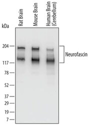
- Western blot analysis of Neurofascin in rat brain tissue, mouse brain tissue, and human brain (cerebellum). Samples were incubated in Neurofascin polyclonal antibody (Product # PA5-47468) using a dilution of 0.1 µg/mL followed by a HRP-conjugated Anti-Chicken IgY secondary antibody. Specific bands were detected for Neurofascin at approximately 140 and186 kDa (as indicated). This experiment was conducted under reducing conditions.
Supportive validation
- Submitted by
- Invitrogen Antibodies (provider)
- Main image

- Experimental details
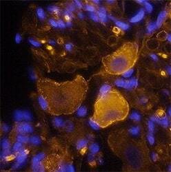
- Immunohistochemical analysis of Neurofascin in perfusion fixed frozen sections of rat brain (dorsal root ganglion). Samples were incubated in Neurofascin polyclonal antibody (Product # PA5-47468) using a dilution of 1.7 µg/mL overnight at 4 °C followed by NorthernLights™ 557-conjugated Anti-Chicken IgY Secondary Antibody (yellow) and counterstained with DAPI (blue). Specific staining was localized to neuronal cell bodies and Schwann cells (perinodal regions).
 Explore
 Validate
 Learn
Learn Western blot