Antibody data
- Antibody Data
- Antigen structure
- References [3]
- Comments [0]
- Validations
- Western blot [1]
- Immunohistochemistry [1]
Submit
Validation data
Reference
Comment
Report error
- Product number
- AF3235 - Provider product page

- Provider
- R&D Systems
- Product name
- Human/Mouse/Rat Neurofascin Antibody
- Antibody type
- Polyclonal
- Description
- Immunogen affinity purified. Detects human, mouse, rat Neurofascin in Western blots and rat Neurofascin in direct ELISAs.
- Reactivity
- Human, Mouse, Rat
- Host
- Chicken/Avian
- Conjugate
- Unconjugated
- Antigen sequence
NP_446361- Isotype
- IgY
- Vial size
- 100 ug
- Concentration
- LYOPH
- Storage
- Use a manual defrost freezer and avoid repeated freeze-thaw cycles. 12 months from date of receipt, -20 to -70 °C as supplied. 1 month, 2 to 8 °C under sterile conditions after reconstitution. 6 months, -20 to -70 °C under sterile conditions after reconstitution.
Submitted references Glial βII Spectrin Contributes to Paranode Formation and Maintenance.
Antibodies against peripheral nerve antigens in chronic inflammatory demyelinating polyradiculoneuropathy.
The paranodal cytoskeleton clusters Na(+) channels at nodes of Ranvier.
Susuki K, Zollinger DR, Chang KJ, Zhang C, Huang CY, Tsai CR, Galiano MR, Liu Y, Benusa SD, Yermakov LM, Griggs RB, Dupree JL, Rasband MN
The Journal of neuroscience : the official journal of the Society for Neuroscience 2018 Jul 4;38(27):6063-6075
The Journal of neuroscience : the official journal of the Society for Neuroscience 2018 Jul 4;38(27):6063-6075
Antibodies against peripheral nerve antigens in chronic inflammatory demyelinating polyradiculoneuropathy.
Querol L, Siles AM, Alba-Rovira R, Jáuregui A, Devaux J, Faivre-Sarrailh C, Araque J, Rojas-Garcia R, Diaz-Manera J, Cortés-Vicente E, Nogales-Gadea G, Navas-Madroñal M, Gallardo E, Illa I
Scientific reports 2017 Oct 31;7(1):14411
Scientific reports 2017 Oct 31;7(1):14411
The paranodal cytoskeleton clusters Na(+) channels at nodes of Ranvier.
Amor V, Zhang C, Vainshtein A, Zhang A, Zollinger DR, Eshed-Eisenbach Y, Brophy PJ, Rasband MN, Peles E
eLife 2017 Jan 30;6
eLife 2017 Jan 30;6
No comments: Submit comment
Supportive validation
- Submitted by
- R&D Systems (provider)
- Main image

- Experimental details
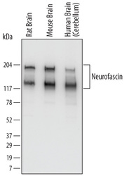
- Detection of Human, Mouse, and Rat Neurofascin by Western Blot. Western blot shows lysates of rat brain tissue, mouse brain tissue, and human brain (cerebellum). PVDF Membrane was probed with 0.1 µg/mL of Chicken Anti-Human/Mouse/Rat Neurofascin Antigen Affinity-purified Polyclonal Antibody (Catalog # AF3235) followed by HRP-conjugated Anti-Chicken IgY Secondary Antibody. Specific bands were detected for Neurofascin at approximately 140 and186 kDa (as indicated). This experiment was conducted under reducing conditions and using Immunoblot Buffer Group 1.
Supportive validation
- Submitted by
- R&D Systems (provider)
- Main image

- Experimental details
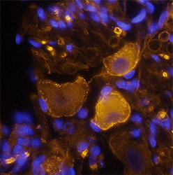
- Neurofascin in Rat Brain. Neurofascin was detected in perfusion fixed frozen sections of rat brain (dorsal root ganglion) using Chicken Anti-Human/Mouse/Rat Neurofascin Antigen Affinity-purified Polyclonal Antibody (Catalog # AF3235) at 1.7 µg/mL overnight at 4 °C. Tissue was stained using the NorthernLights™ 557-conjugated Anti-Chicken IgY Secondary Antibody (yellow; Catalog # NL016) and counterstained with DAPI (blue). Specific staining was localized to neuronal cell bodies and Schwann cells (perinodal regions). View our protocol for Fluorescent IHC Staining of Frozen Tissue Sections.
 Explore
 Validate
 Learn
Learn Western blot