HPA008213
antibody from Atlas Antibodies
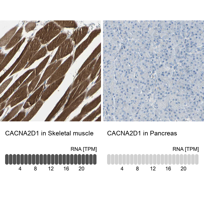
Targeting: CACNA2D1
CACNA2, CACNL2A, LINC01112, lncRNA-N3, MHS3
Antibody data
- Antibody Data
- Antigen structure
- References [1]
- Comments [0]
- Validations
- Western blot [1]
- Immunohistochemistry [14]
Submit
Validation data
Reference
Comment
Report error
- Product number
- HPA008213 - Provider product page
- Provider
- Atlas Antibodies
- Proper citation
- Atlas Antibodies Cat#HPA008213, RRID:AB_1233654
- Product name
- Anti-CACNA2D1
- Antibody type
- Polyclonal
- Reactivity
- Human
- Host
- Rabbit
- Conjugate
- Unconjugated
- Antigen sequence
QPKNPKSQEPVTLDFLDAELENDIKVEIRNKMIDG
ESGEKTFRTLVKSQDERYIDKGNRTYTWTPVNGTD
YSLALVLPTYSFYYIKAKLEETITQARSKKGKMKD
SETLKPDNFEESGYTFIAPRDYCNDLKISDNNTEF
L- Isotype
- IgG
- Vial size
- 100 µl
- Storage
- Store at +4°C for short term storage. Long time storage is recommended at -20°C.
Submitted references PBX3 is targeted by multiple miRNAs and is essential for liver tumour-initiating cells
Han H, Du Y, Zhao W, Li S, Chen D, Zhang J, Liu J, Suo Z, Bian X, Xing B, Zhang Z
Nature Communications 2015 September;6
No comments: Submit comment
Supportive validation
- Submitted by
- Atlas Antibodies (provider)
- Enhanced method
- Orthogonal validation
- Main image
- Experimental details
- Western blot analysis in human cell line U-2 OS and human cell line SK-MEL-30.
Enhanced validation
Enhanced validation
Supportive validation
- Submitted by
- Atlas Antibodies (provider)
- Enhanced method
- Orthogonal validation
- Main image
- Experimental details
- Immunohistochemistry analysis in human duodenum and pancreas tissues using HPA008213 antibody. Corresponding CACNA2D1 RNA-seq data are presented for the same tissues.
- Sample type
- HUMAN
Enhanced validation
- Submitted by
- Atlas Antibodies (provider)
- Enhanced method
- Independent antibody validation
- Main image
- Experimental details
- Immunohistochemical staining of human cerebral cortex, kidney, liver and skeletal muscle using Anti-CACNA2D1 antibody HPA008213 (A) shows similar protein distribution across tissues to independent antibody HPA008621 (B).
Supportive validation
- Submitted by
- Atlas Antibodies (provider)
- Main image
- Experimental details
- Immunohistochemical staining of human skeletal muscle shows strong cytoplasmic positivity in myocytes.
- Submitted by
- Atlas Antibodies (provider)
- Main image
- Experimental details
- Immunohistochemical staining of human skeletal muscle shows strong cytoplasmic positivity in myocytes.
- Sample type
- HUMAN
- Submitted by
- Atlas Antibodies (provider)
- Main image
- Experimental details
- Immunohistochemical staining of human cerebral cortex shows moderate positivity.
- Sample type
- HUMAN
- Submitted by
- Atlas Antibodies (provider)
- Main image
- Experimental details
- Immunohistochemical staining of human cerebellum shows moderate positivity.
- Sample type
- HUMAN
- Submitted by
- Atlas Antibodies (provider)
- Main image
- Experimental details
- Immunohistochemical staining of human pancreas shows no positivity in exocrine glandular cells as expected.
- Sample type
- HUMAN
- Submitted by
- Atlas Antibodies (provider)
- Main image
- Experimental details
- Immunohistochemical staining of human small intestine shows moderate to strong membranous positivity in glandular cells.
- Sample type
- HUMAN
- Submitted by
- Atlas Antibodies (provider)
- Main image
- Experimental details
- Immunohistochemical staining of human duodenum shows moderate to strong membranous positivity in glandular cells.
- Sample type
- HUMAN
- Submitted by
- Atlas Antibodies (provider)
- Main image
- Experimental details
- Immunohistochemical staining of human pancreas shows no positivity in exocrine glandular cells as expected.
- Sample type
- HUMAN
- Submitted by
- Atlas Antibodies (provider)
- Main image
- Experimental details
- Immunohistochemical staining of human kidney using Anti-CACNA2D1 antibody HPA008213.
- Sample type
- HUMAN
- Submitted by
- Atlas Antibodies (provider)
- Main image
- Experimental details
- Immunohistochemical staining of human liver using Anti-CACNA2D1 antibody HPA008213.
- Sample type
- HUMAN
- Submitted by
- Atlas Antibodies (provider)
- Main image
- Experimental details
- Immunohistochemical staining of human skeletal muscle using Anti-CACNA2D1 antibody HPA008213.
- Sample type
- HUMAN
- Submitted by
- Atlas Antibodies (provider)
- Main image
- Experimental details
- Immunohistochemical staining of human cerebral cortex using Anti-CACNA2D1 antibody HPA008213.
- Sample type
- HUMAN