Explore
 Validate
 Learn
Learn Western blot
Antibody data
- Antibody Data
- Antigen structure
- References [0]
- Comments [0]
- Validations
- Western blot [1]
- Immunohistochemistry [1]
- Other assay [1]
Submit
Validation data
Reference
Comment
Report error
- Product number
- PA5-19228 - Provider product page

- Provider
- Invitrogen Antibodies
- Product name
- Defensin 1/3 Polyclonal Antibody
- Antibody type
- Polyclonal
- Antigen
- Synthetic peptide
- Description
- This antibody is tested in Peptide ELISA: antibody detection limit dilution 128,000.
- Reactivity
- Human
- Host
- Goat
- Isotype
- IgG
- Vial size
- 100 μg
- Concentration
- 0.5 mg/mL
- Storage
- -20°C, Avoid Freeze/Thaw Cycles
No comments: Submit comment
Supportive validation
- Submitted by
- Invitrogen Antibodies (provider)
- Main image
- Experimental details
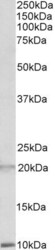
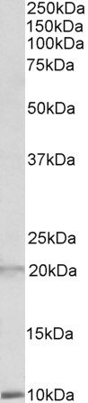
- Western blot analysis of Defensin 1/3 using Defensin 1/3 Polyclonal Antibody (Product # PA5-19228) (2 µg/mL) in staining of Human Spleen lysate (35 µg protein in RIPA buffer). Detected by chemiluminescence.
Supportive validation
- Submitted by
- Invitrogen Antibodies (provider)
- Main image
- Experimental details
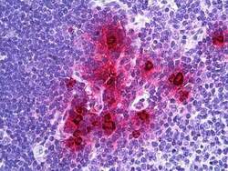
- Immunohistochemistry analysis of Defensin 1/3 in human thymus, myeloid cells. Samples were incubated with Defensin 1/3 polyclonal antibody (Product # PA5-19228) using a dilution of 3.75 µg/mL. Formalin-fixed, paraffin-embedded tissue after heat-induced antigen retrieval.

Supportive validation
- Submitted by
- Invitrogen Antibodies (provider)
- Main image

- Experimental details
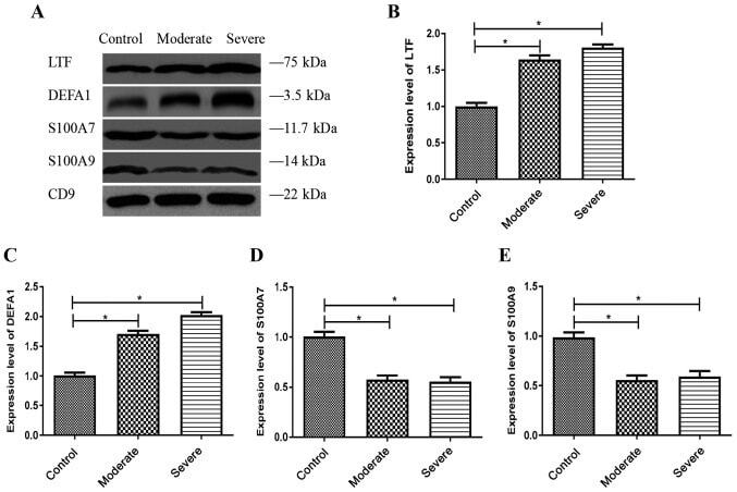
- Figure 6. Verification of differentially expressed proteins using western blotting. (A) Representative western blots demonstrate the expression levels of LTF, DEFA1, S100A7 and S100A9 in the MV/E isolated from the CSF. Histogram data shows the densitometric semi-quantification (mean +- SD) of (B) LTF, (C) DEFA1, (D) S100A7 and (E) S100A9 expression levels. Protein densitometric semi-quantification was normalized to the density of CD9 protein. CD9 is an exosome surface marker. *P