Explore
 Validate
 Learn
Learn Western blot
Antibody data
- Antibody Data
- Antigen structure
- References [2]
- Comments [0]
- Validations
- Western blot [2]
- Immunocytochemistry [3]
- Immunohistochemistry [2]
- Other assay [1]
Submit
Validation data
Reference
Comment
Report error
- Product number
- PA1-104 - Provider product page

- Provider
- Invitrogen Antibodies
- Product name
- GATA6 Polyclonal Antibody
- Antibody type
- Polyclonal
- Antigen
- Synthetic peptide
- Reactivity
- Human, Mouse
- Host
- Rabbit
- Isotype
- IgG
- Vial size
- 100 μg
- Concentration
- 1 mg/mL
- Storage
- -20°C
Submitted references Construction of a mammalian embryo model from stem cells organized by a morphogen signalling centre.
Reproducible differentiation and characterization of neurons from mouse embryonic stem cells.
Xu PF, Borges RM, Fillatre J, de Oliveira-Melo M, Cheng T, Thisse B, Thisse C
Nature communications 2021 Jun 2;12(1):3277
Nature communications 2021 Jun 2;12(1):3277
Reproducible differentiation and characterization of neurons from mouse embryonic stem cells.
Saxena S, Choudhury S, Mohan KN
MethodsX 2020;7:101073
MethodsX 2020;7:101073
No comments: Submit comment
Supportive validation
- Submitted by
- Invitrogen Antibodies (provider)
- Main image
- Experimental details
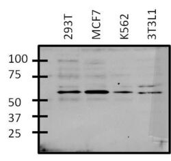
- Western blot analysis of GATA6 was performed by loading 25 µg of various whole cell lysate onto a 4-20% Tris-HCl polyacrylamide gel. Proteins were transferred to a PVDF membrane and blocked with 5% BSA/TBST for at least 1 hour. Membranes were then probed with a rabbit polyclonal antibody recognizing GATA6 (Product # PA1-104) at a dilution of 1:1000 overnight at 4°C on a rocking platform. Membranes were then washed in TBS-0.1%Tween 20 and probed with a goat anti-rabbit-HRP secondary antibody (Product # 31460) at a dilution of 1:15,000 for at least one hour. Membranes were washed and chemiluminescent detection was performed using SuperSignal West Pico (Product # 34080).

- Submitted by
- Invitrogen Antibodies (provider)
- Main image

- Experimental details
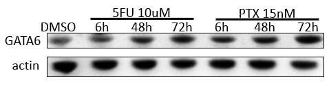
- Western blot analysis of GATA6 was performed by loading 100 ng of human BMSC whole cell lysates treated with either 5FU or PTX as indicated in RIPA Lysis and Extraction buffer (Product # 89900) and Page Ruler Plus Protein Ladder (Product # 26619) onto a 30% Acrylamide/Bis gel. Proteins were transferred to membrane and blocked in 5% milk for one hour at room temperature. GATA6 was detected using a GATA6 polyclonal antibody (Product # PA1-104) at a dilution of 1:1,000 in 5% milk overnight at 4°C on a rocking platform, followed by a goat anti-rabit IgG-HRP secondary antibody at a dilution of 1:5,000 for one hour. Chemiluminescent detection was performed using SuperSignal West Dura (Product # 34076) and the myECL Imager (Product # 62236). Data courtesy of Wei Xu’s laboratory at the University of Wisconsin.
Supportive validation
- Submitted by
- Invitrogen Antibodies (provider)
- Main image

- Experimental details
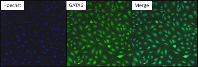
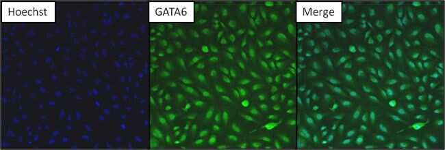
- Immunofluorescent analysis of GATA6 (green) in HeLa cells. Formalin fixed cells were permeabilized with 0.1% Triton X-100 in TBS for 10 minutes at room temperature. Cells were then blocked with 5% normal goat serum (Product # 31873) for 15 minutes at room temperature. Cells were then probed with a rabbit polyclonal antibody recognizing GATA6 (Product # PA1-104) at a dilution of 1:200 for at least 1 hour at 37°C. Cells were then washed with PBS and incubated with DyLight 488 goat-anti-rabbit secondary antibody at a dilution of 1:200 for 30 minutes at room temperature. Nuclei (blue) were stained with Hoechst 33342 dye (Product # 62249). Images were taken on a Thermo Scientific ArrayScan at 10X magnification.
- Submitted by
- Invitrogen Antibodies (provider)
- Main image

- Experimental details
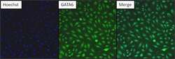
- Immunofluorescent analysis of GATA6 (green) in HeLa cells. Formalin fixed cells were permeabilized with 0.1% Triton X-100 in TBS for 10 minutes at room temperature. Cells were then blocked with 5% normal goat serum (Product # 31873) for 15 minutes at room temperature. Cells were then probed with a rabbit polyclonal antibody recognizing GATA6 (Product # PA1-104) at a dilution of 1:200 for at least 1 hour at 37°C. Cells were then washed with PBS and incubated with DyLight 488 goat-anti-rabbit secondary antibody at a dilution of 1:200 for 30 minutes at room temperature. Nuclei (blue) were stained with Hoechst 33342 dye (Product # 62249). Images were taken on a Thermo Scientific ArrayScan at 10X magnification.
- Submitted by
- Invitrogen Antibodies (provider)
- Main image
- Experimental details
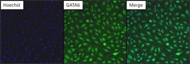
- Immunofluorescent analysis of GATA6 (green) in HeLa cells. Formalin fixed cells were permeabilized with 0.1% Triton X-100 in TBS for 10 minutes at room temperature. Cells were then blocked with 5% normal goat serum (Product # 31873) for 15 minutes at room temperature. Cells were then probed with a rabbit polyclonal antibody recognizing GATA6 (Product # PA1-104) at a dilution of 1:200 for at least 1 hour at 37°C. Cells were then washed with PBS and incubated with DyLight 488 goat-anti-rabbit secondary antibody at a dilution of 1:200 for 30 minutes at room temperature. Nuclei (blue) were stained with Hoechst 33342 dye (Product # 62249). Images were taken on a Thermo Scientific ArrayScan at 10X magnification.
Supportive validation
- Submitted by
- Invitrogen Antibodies (provider)
- Main image
- Experimental details
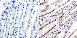
- Immunohistochemistry was performed on human stomach tissue. To expose target protein, antigen was retreived using 10mM sodium citrate followed by microwave treatment for 8-15 minutes. Endogenous peroxidases were blocked in 3% H202-methanol for 15 minutes and tissues were blocked in 3% BSA-PBS for 30 minutes at room temperature. Cells were probed with a GATA6 Rabbit polyclonal antibody (Product # PA1-104) at a dilution of 1:100 overnight in a humidified chamber. Tissues were washed in PBST and detection was performed using a secondary antibody conjugated to HRP. DAB staining buffer was applied and tissues were counterstained with hematoxylin and prepped for mounting. Images were taken at 40X magnification.

- Submitted by
- Invitrogen Antibodies (provider)
- Main image

- Experimental details
- Immunohistochemistry was performed on human stomach tissue. To expose target protein, antigen was retreived using 10mM sodium citrate followed by microwave treatment for 8-15 minutes. Endogenous peroxidases were blocked in 3% H202-methanol for 15 minutes and tissues were blocked in 3% BSA-PBS for 30 minutes at room temperature. Cells were probed with a GATA6 Rabbit polyclonal antibody (Product # PA1-104) at a dilution of 1:100 overnight in a humidified chamber. Tissues were washed in PBST and detection was performed using a secondary antibody conjugated to HRP. DAB staining buffer was applied and tissues were counterstained with hematoxylin and prepped for mounting. Images were taken at 40X magnification.
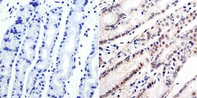
Supportive validation
- Submitted by
- Invitrogen Antibodies (provider)
- Main image

- Experimental details
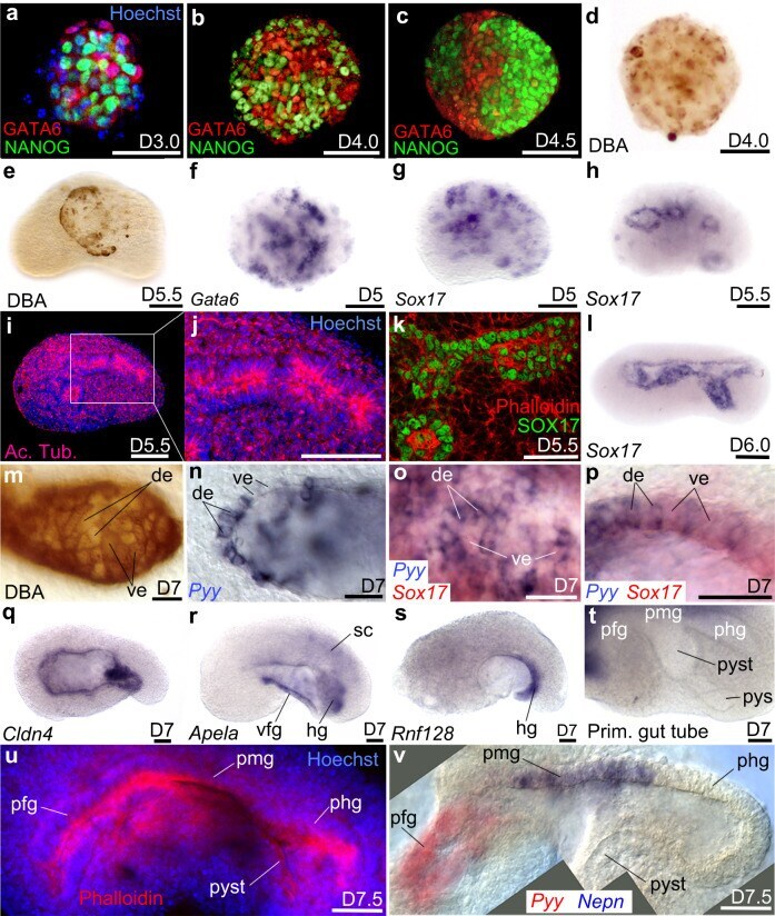
- Fig. 3 Endoderm germ layer and formation of the embryoid gut tube. a - c Immunodetection of NANOG and GATA6 from D3.0 to D4.5. d , e DBA labelling of visceral endoderm (ve) at d D4.0 and e at the surface of the embryoid at D5.5. f , g Clumps of endoderm cells expressing f Gata6 or g Sox17 . At D5.5 h Sox17 was expressed around small cavities bordered i , j by a polarized epithelium positive for Acetylated Tubulin (Ac. Tub). k Immunodetection of SOX17 identifies the endodermal-like epithelium. l In situ hybridization for Sox17 at D6.0 after the merging of small cavities into a single one. m Mosaic of DBA(+) (ve cells) and DBA(-) cells (definitive endoderm: de) in the endoderm epithelium. n Patchy expression of Pyy (de marker). o , p Expression of Sox17 (red) and Pyy (blue) showing that the endoderm epithelium was a mosaic of de and ve. o Superficial view of the epithelium. p Optical cross-section of the epithelium at higher magnification. q - s In situ hybridization for q Cldn4 in the whole gut epithelium, r Apela in the ventral foregut (vfg) and hindgut (hg), s Rnf128 in the hg. t Folding of the primitive-like gut showing a presumptive fg (pfg) followed dorso-posteriorly by likely presumptive midgut (pmg), presumptive hindgut (phg), presumptive yolk stalk (pyst) and presumptive yolk sac (pys). u Phalloidin (red) and Hoechst (nuclei, blue) labelling of primitive gut tube. v Combined images of a double colour in situ hybridization of pgt at D7.5 showing expression of Pyy (red)