Explore
 Validate
 Learn
Learn Western blot
 Immunocytochemistry
Antibody data
- Antibody Data
- Antigen structure
- References [2]
- Comments [0]
- Validations
- Immunocytochemistry [3]
- Immunohistochemistry [1]
- Other assay [1]
Submit
Validation data
Reference
Comment
Report error
- Product number
- PA1-104X - Provider product page

- Provider
- Invitrogen Antibodies
- Product name
- GATA6 Polyclonal Antibody
- Antibody type
- Polyclonal
- Antigen
- Synthetic peptide
- Reactivity
- Human, Mouse
- Host
- Rabbit
- Isotype
- IgG
- Vial size
- 20 μL
- Concentration
- 1 mg/mL
- Storage
- -20°C
Submitted references Construction of a mammalian embryo model from stem cells organized by a morphogen signalling centre.
Reproducible differentiation and characterization of neurons from mouse embryonic stem cells.
Xu PF, Borges RM, Fillatre J, de Oliveira-Melo M, Cheng T, Thisse B, Thisse C
Nature communications 2021 Jun 2;12(1):3277
Nature communications 2021 Jun 2;12(1):3277
Reproducible differentiation and characterization of neurons from mouse embryonic stem cells.
Saxena S, Choudhury S, Mohan KN
MethodsX 2020;7:101073
MethodsX 2020;7:101073
No comments: Submit comment
Supportive validation
- Submitted by
- Invitrogen Antibodies (provider)
- Main image

- Experimental details
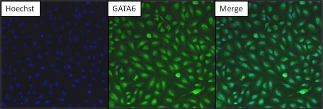
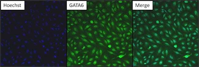
- Immunofluorescent analysis of GATA6 (green) in HeLa cells. Formalin fixed cells were permeabilized with 0.1% Triton X-100 in TBS for 10 minutes at room temperature. Cells were then blocked with 5% normal goat serum (Product # 31873) for 15 minutes at room temperature. Cells were then probed with a rabbit polyclonal antibody recognizing GATA6 (Product # PA1-104) at a dilution of 1:200 for at least 1 hour at 37°C. Cells were then washed with PBS and incubated with DyLight 488 goat-anti-rabbit secondary antibody at a dilution of 1:200 for 30 minutes at room temperature. Nuclei (blue) were stained with Hoechst 33342 dye (Product # 62249). Images were taken on a Thermo Scientific ArrayScan at 10X magnification.
- Submitted by
- Invitrogen Antibodies (provider)
- Main image

- Experimental details
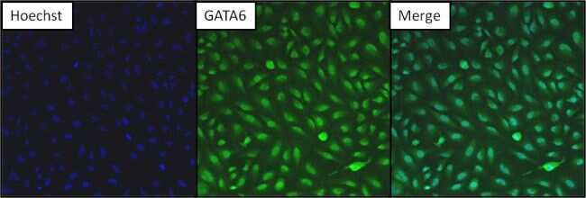
- Immunofluorescent analysis of GATA6 (green) in HeLa cells. Formalin fixed cells were permeabilized with 0.1% Triton X-100 in TBS for 10 minutes at room temperature. Cells were then blocked with 5% normal goat serum (Product # 31873) for 15 minutes at room temperature. Cells were then probed with a rabbit polyclonal antibody recognizing GATA6 (Product # PA1-104) at a dilution of 1:200 for at least 1 hour at 37°C. Cells were then washed with PBS and incubated with DyLight 488 goat-anti-rabbit secondary antibody at a dilution of 1:200 for 30 minutes at room temperature. Nuclei (blue) were stained with Hoechst 33342 dye (Product # 62249). Images were taken on a Thermo Scientific ArrayScan at 10X magnification.
- Submitted by
- Invitrogen Antibodies (provider)
- Main image
- Experimental details
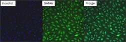
- Immunofluorescent analysis of GATA6 (green) in HeLa cells. Formalin fixed cells were permeabilized with 0.1% Triton X-100 in TBS for 10 minutes at room temperature. Cells were then blocked with 5% normal goat serum (Product # 31873) for 15 minutes at room temperature. Cells were then probed with a rabbit polyclonal antibody recognizing GATA6 (Product # PA1-104) at a dilution of 1:200 for at least 1 hour at 37°C. Cells were then washed with PBS and incubated with DyLight 488 goat-anti-rabbit secondary antibody at a dilution of 1:200 for 30 minutes at room temperature. Nuclei (blue) were stained with Hoechst 33342 dye (Product # 62249). Images were taken on a Thermo Scientific ArrayScan at 10X magnification.
Supportive validation
- Submitted by
- Invitrogen Antibodies (provider)
- Main image
- Experimental details
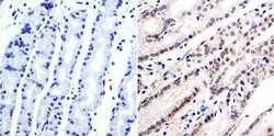
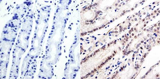
- Immunohistochemistry was performed on human stomach tissue. To expose target protein, antigen was retreived using 10mM sodium citrate followed by microwave treatment for 8-15 minutes. Endogenous peroxidases were blocked in 3% H202-methanol for 15 minutes and tissues were blocked in 3% BSA-PBS for 30 minutes at room temperature. Cells were probed with a GATA6 Rabbit polyclonal antibody (Product # PA1-104) at a dilution of 1:100 overnight in a humidified chamber. Tissues were washed in PBST and detection was performed using a secondary antibody conjugated to HRP. DAB staining buffer was applied and tissues were counterstained with hematoxylin and prepped for mounting. Images were taken at 40X magnification.
Supportive validation
- Submitted by
- Invitrogen Antibodies (provider)
- Main image

- Experimental details
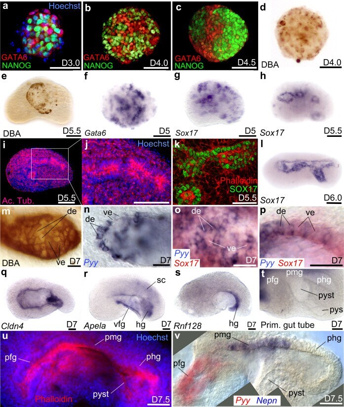
- Fig. 3 Endoderm germ layer and formation of the embryoid gut tube. a - c Immunodetection of NANOG and GATA6 from D3.0 to D4.5. d , e DBA labelling of visceral endoderm (ve) at d D4.0 and e at the surface of the embryoid at D5.5. f , g Clumps of endoderm cells expressing f Gata6 or g Sox17 . At D5.5 h Sox17 was expressed around small cavities bordered i , j by a polarized epithelium positive for Acetylated Tubulin (Ac. Tub). k Immunodetection of SOX17 identifies the endodermal-like epithelium. l In situ hybridization for Sox17 at D6.0 after the merging of small cavities into a single one. m Mosaic of DBA(+) (ve cells) and DBA(-) cells (definitive endoderm: de) in the endoderm epithelium. n Patchy expression of Pyy (de marker). o , p Expression of Sox17 (red) and Pyy (blue) showing that the endoderm epithelium was a mosaic of de and ve. o Superficial view of the epithelium. p Optical cross-section of the epithelium at higher magnification. q - s In situ hybridization for q Cldn4 in the whole gut epithelium, r Apela in the ventral foregut (vfg) and hindgut (hg), s Rnf128 in the hg. t Folding of the primitive-like gut showing a presumptive fg (pfg) followed dorso-posteriorly by likely presumptive midgut (pmg), presumptive hindgut (phg), presumptive yolk stalk (pyst) and presumptive yolk sac (pys). u Phalloidin (red) and Hoechst (nuclei, blue) labelling of primitive gut tube. v Combined images of a double colour in situ hybridization of pgt at D7.5 showing expression of Pyy (red)