 Explore
 Validate
 Learn
LearnMA5-41051
antibody from Invitrogen Antibodies
Targeting: HSPA4
HS24/P52, HSPH2
 Western blot
 ELISA
ELISA Immunocytochemistry Immunoprecipitation Immunohistochemistry Flow cytometry Other assay
Antibody data
- Antibody Data
- Antigen structure
- References [0]
- Comments [0]
- Validations
- Immunocytochemistry [3]
- Immunoprecipitation [2]
- Immunohistochemistry [4]
- Flow cytometry [3]
- Other assay [2]
Submit
Validation data
Reference
Comment
Report error
- Product number
- MA5-41051 - Provider product page

- Provider
- Invitrogen Antibodies
- Product name
- HSP70 Recombinant Rabbit Monoclonal Antibody (001)
- Antibody type
- Monoclonal
- Antigen
- Recombinant full-length protein
- Reactivity
- Human
- Host
- Rabbit
- Isotype
- IgG
- Antibody clone number
- 001
- Vial size
- 100 μL
- Concentration
- 1 mg/mL
- Storage
- -20°C, Avoid Freeze/Thaw Cycles
No comments: Submit comment
Supportive validation
- Submitted by
- Invitrogen Antibodies (provider)
- Main image
- Experimental details
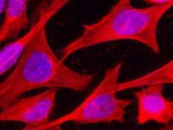
- Immunofluorescence staining of Human HSP70 in Hela cells. Cells were fixed with 4% PFA, permeabilzed with 0.3% Triton X-100 in PBS, blocked with 10% serum, and incubated with HSP70 Recombinant Rabbit Monoclonal Antibody (001) (Product # MA5-41051, 1:60) at 37°C 1 hour. Then cells were stained with the Alexa Fluor® 594-conjugated goat Anti-rabbit IgG secondary antibody (red) and counterstained with DAPI (blue). Positive staining was localized to cytoplasm.

- Submitted by
- Invitrogen Antibodies (provider)
- Main image

- Experimental details
- Immunocytochemistry analysis of HSP70 in HeLa cells. Samples were treated with 4% PFA, permeabilzed with 0.3% Triton X-100 in PBS, blocked with 10% serum, incubated with monoclonal antibody (Product # MA5-41051) with a dilution of 1:60 (37°C 1 hour), followed by Alexa Fluor 594-conjugated goat Anti-rabbit IgG secondary antibody (red) and DAPI.

- Submitted by
- Invitrogen Antibodies (provider)
- Main image

- Experimental details
- Immunofluorescence staining of Human HSP70 in Hela cells. Cells were fixed with 4% PFA, permeabilzed with 0.3% Triton X-100 in PBS, blocked with 10% serum, and incubated with HSP70 Recombinant Rabbit Monoclonal Antibody (001) (Product # MA5-41051, 1:60) at 37°C 1 hour. Then cells were stained with the Alexa Fluor® 594-conjugated goat Anti-rabbit IgG secondary antibody (red) and counterstained with DAPI (blue). Positive staining was localized to cytoplasm.

Supportive validation
- Submitted by
- Invitrogen Antibodies (provider)
- Main image

- Experimental details
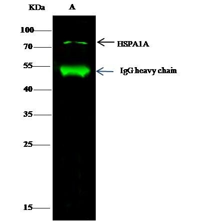
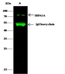
- HSP70 Immunoprecipitation using: Lane A: 0.5 mg Hela Whole Cell Lysate 2 µL with HSP70 Recombinant Rabbit Monoclonal Antibody (001) (Product # MA5-41051) and 15 µL of 50 % Protein G agarose. Primary antibody: HSP70 Recombinant Rabbit Monoclonal Antibody (001), at 1:200 dilution. Secondary antibody: Dylight 800-labeled antibody to rabbit IgG (H+L), at 1:5,000 dilution. Developed using the Odyssey technique. Performed under reducing conditions. Predicted band size: 70 kDa. Observed band size: 70 kDa.
- Submitted by
- Invitrogen Antibodies (provider)
- Main image

- Experimental details
- Immunoprecipitation of HSP70 in Lane A: 0.5 mg Hela Whole Cell Lysate. Samples were treated with 15 μl of 50 % Protein G agarose, incubated with monoclonal antibody (Product # MA5-41051) with a dilution of 1:200 , followed by Dylight 800-labeled antibody to rabbit IgG (H+L) using a dilution of 1:5,000. Assay was performed under reducing conditions. Predicted band size: 70 kDa, Observed band size: 70 kDa .

Supportive validation
- Submitted by
- Invitrogen Antibodies (provider)
- Main image
- Experimental details
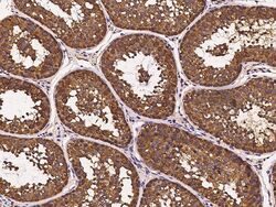
- Immunohistochemistry (Paraffin) of HSP70 in cynomolgus macaque testis. Samples were incubated with monoclonal antibody (Product # MA5-41051) with a dilution of 1:1,000.

- Submitted by
- Invitrogen Antibodies (provider)
- Main image
- Experimental details
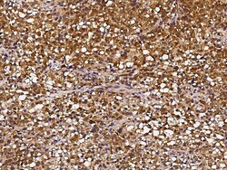
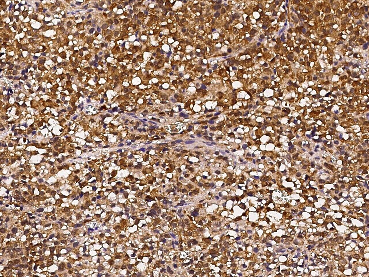
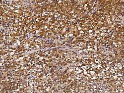
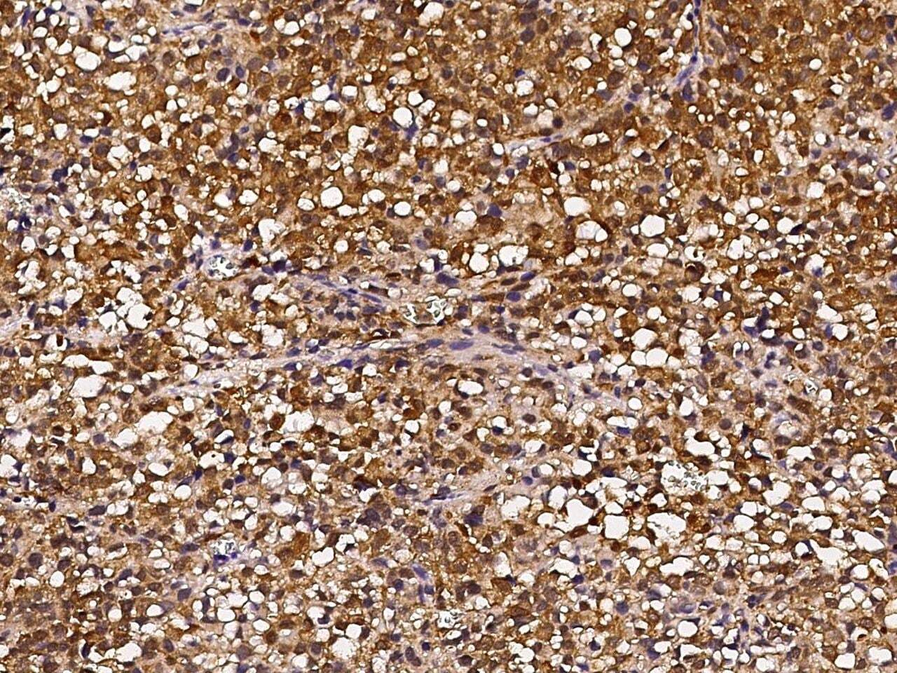
- Immunohistochemical staining of HSP70 in human hepatoma with HSP70 Recombinant Rabbit Monoclonal Antibody (001) (Product # MA5-41051, 1:1,000, formalin-fixed paraffin embedded sections).
- Submitted by
- Invitrogen Antibodies (provider)
- Main image
- Experimental details
- Immunohistochemistry (Paraffin) of HSP70 in human hepatoma. Samples were incubated with monoclonal antibody (Product # MA5-41051) with a dilution of 1:1,000.
- Submitted by
- Invitrogen Antibodies (provider)
- Main image

- Experimental details
- Immunohistochemical staining of HSP70 in cynomolgus macaque testis with HSP70 Recombinant Rabbit Monoclonal Antibody (001) (Product # MA5-41051, 1:1,000, formalin-fixed paraffin embedded sections).

Supportive validation
- Submitted by
- Invitrogen Antibodies (provider)
- Main image

- Experimental details
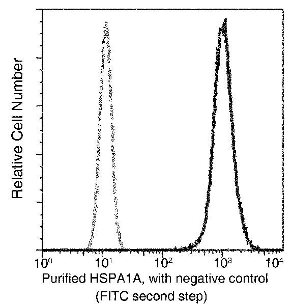
- Flow cytometric analysis of Human HSP70 expression on HeLa cells. The cells were treated according to manufacturer’s manual, stained with HSP70 Recombinant Rabbit Monoclonal Antibody (001) (Product # MA5-41051), then a FITC-conjugated Secondary antibody. The fluorescence histograms were derived from gated events with the forward and side light-scatter characteristics of intact cells.

- Submitted by
- Invitrogen Antibodies (provider)
- Main image

- Experimental details
- Flow Cytometry of HSP70 in HeLa cells. Samples were incubated with monoclonal antibody (Product # MA5-41051) followed by FITC.

- Submitted by
- Invitrogen Antibodies (provider)
- Main image

- Experimental details
- Flow cytometric analysis of Human HSP70 expression on HeLa cells. The cells were treated according to manufacturer’s manual, stained with HSP70 Recombinant Rabbit Monoclonal Antibody (001) (Product # MA5-41051), then a FITC-conjugated Secondary antibody. The fluorescence histograms were derived from gated events with the forward and side light-scatter characteristics of intact cells.
Supportive validation
- Submitted by
- Invitrogen Antibodies (provider)
- Main image
- Experimental details
- Immunoprecipitation of HSP70 in Lane A: 0.5 mg Hela Whole Cell Lysate. Samples were treated with 15 μl of 50 % Protein G agarose, incubated with monoclonal antibody (Product # MA5-41051) with a dilution of 1:200 , followed by Dylight 800-labeled antibody to rabbit IgG (H+L) using a dilution of 1:5,000. Assay was performed under reducing conditions. Predicted band size: 70 kDa, Observed band size: 70 kDa .

- Submitted by
- Invitrogen Antibodies (provider)
- Main image

- Experimental details
- HSP70 Immunoprecipitation using: Lane A: 0.5 mg Hela Whole Cell Lysate 2 µL with HSP70 Recombinant Rabbit Monoclonal Antibody (001) (Product # MA5-41051) and 15 µL of 50 % Protein G agarose. Primary antibody: HSP70 Recombinant Rabbit Monoclonal Antibody (001), at 1:200 dilution. Secondary antibody: Dylight 800-labeled antibody to rabbit IgG (H+L), at 1:5,000 dilution. Developed using the Odyssey technique. Performed under reducing conditions. Predicted band size: 70 kDa. Observed band size: 70 kDa.
