Explore
 Validate
 Learn
Learn Western blot
 ELISA
ELISAAntibody data
- Antibody Data
- Antigen structure
- References [1]
- Comments [0]
- Validations
- Western blot [2]
- Immunocytochemistry [2]
- Immunohistochemistry [1]
Submit
Validation data
Reference
Comment
Report error
- Product number
- MA5-15578 - Provider product page

- Provider
- Invitrogen Antibodies
- Product name
- HSPA4 Monoclonal Antibody (5A6)
- Antibody type
- Monoclonal
- Antigen
- Purifed from natural sources
- Description
- MA5-15578 targets HSP70 in indirect ELISA, IF, IHC, and WB applications and shows reactivity with Human samples. The MA5-15578 immunogen is purified recombinant fragment of human HSP70 expressed in E. Coli. MA5-15578 detects HSP70 which has a predicted molecular weight of approximately 70kDa.
- Reactivity
- Human, Mouse
- Host
- Mouse
- Isotype
- IgG
- Antibody clone number
- 5A6
- Vial size
- 100 µL
- Concentration
- 1.0 mg/mL
- Storage
- Store at 4°C short term. For long term storage, store at -20°C, avoiding freeze/thaw cycles.
Submitted references Heteromeric complexes of heat shock protein 70 (HSP70) family members, including Hsp70B', in differentiated human neuronal cells.
Chow AM, Mok P, Xiao D, Khalouei S, Brown IR
Cell stress & chaperones 2010 Sep;15(5):545-53
Cell stress & chaperones 2010 Sep;15(5):545-53
No comments: Submit comment
Supportive validation
- Submitted by
- Invitrogen Antibodies (provider)
- Main image
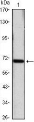
- Experimental details
- Western blot analysis of HSPA4/Heat Shock 70 Protein 4 using HSPA4/Heat Shock 70 Protein 4 monoclonal antibody (Product # MA5-15578) in HeLa (1) cell lysate.
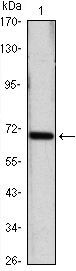
- Submitted by
- Invitrogen Antibodies (provider)
- Main image
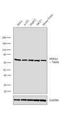
- Experimental details
- Western blot analysis was performed on whole cell extracts (30 µg lysate) of HeLa (Lane 1), A-431 (Lane 2), HepG2 (Lane 3), MCF7 (Lane 4) and Mouse Testis (Lane 5). The blot was probed with Anti-HSPA4 Monoclonal Antibody (Product # MA5-15578, 1:2000 dilution) and detected by chemiluminescence using Goat anti-Mouse IgG (H+L) Superclonal™ Secondary Antibody, HRP conjugate (Product # A28177, 1:4000 dilution). A 70kDa band corresponding to HSPA4 was observed across all cell lines and tissue lysates tested.

Supportive validation
- Submitted by
- Invitrogen Antibodies (provider)
- Main image
- Experimental details
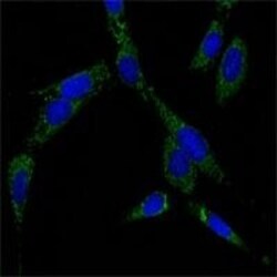
- Immunofluorescence analysis of NIH/3T3 cells using HSPA4/Heat Shock 70 Protein 4 monoclonal antibody (Product # MA5-15578) (Green). Blue: DRAQ5 fluorescent DNA dye.

- Submitted by
- Invitrogen Antibodies (provider)
- Main image

- Experimental details
- Immunofluorescence analysis of HSPA4 was performed using 70% confluent log phase HeLa cells. The cells were fixed with 4% paraformaldehyde for 10 minutes, permeabilized with 0.1% Triton™ X-100 for 15 minutes, and blocked with 1% BSA for 1 hour at room temperature. The cells were labeled with HSPA4 Monoclonal Antibody (5A6) (Product # MA5-15578) at 1:100 dilution in 0.1% BSA, incubated at 4 degree Celsius overnight and then labeled with Goat anti-Mouse IgG (H+L) Superclonal™ Secondary Antibody, Alexa Fluor® 488 conjugate (Product # A28175) at a dilution of 1:2000 for 45 minutes at room temperature (Panel a: green). Nuclei (Panel b: blue) were stained with ProLong™ Diamond Antifade Mountant with DAPI (Product # P36962). F-actin (Panel c: red) was stained with Rhodamine Phalloidin (Product # R415). Panel d represents the merged image showing nucleus and cytoplasm localization. Panel e represents control cells with no primary antibody to assess background. The images were captured at 60X magnification.

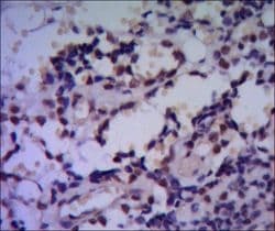
Supportive validation
- Submitted by
- Invitrogen Antibodies (provider)
- Main image

- Experimental details
- Immunohistochemical analysis of paraffin-embedded human breast cancer using HSPA4/Heat Shock 70 Protein 4 monoclonal antibody (Product # MA5-15578) followed with DAB staining.