Explore
 Validate
 Learn
Learn ELISA
ELISA Immunohistochemistry
Antibody data
- Antibody Data
- Antigen structure
- References [4]
- Comments [0]
- Validations
- Immunohistochemistry [1]
- Other assay [1]
Submit
Validation data
Reference
Comment
Report error
- Product number
- 14-9762-82 - Provider product page

- Provider
- Invitrogen Antibodies
- Product name
- Angiogenin Monoclonal Antibody (26-2F), eBioscience™
- Antibody type
- Monoclonal
- Antigen
- Other
- Description
- Description: The monoclonal antibody 26-2F recognizes human angiogenin. Angiogenin is a secreted ribonuclease (RNASE)-family member and is important for the formation of new blood vessels as well as the stimulation of cell proliferation and survival. Cellular stress signals direct angiogenin to stress granules where angiogenin functions by hydrolyzing cellular tRNA resulting in modulation of protein translation and promotion of cell survival. Cytoplasmic angiogenin can bind to actin, forming complexes that activate protease cascades which can degrade laminin and fibronectin allowing endothelial cells to penetrate the basement membrane and migrate. In cancer cells, growth signals direct angiogenin translocation to the nucleus where binding of DNA promotor regions stimulates rRNA transcription resulting in a downstream increase in protein synthesis driving cell growth and survival. Angiogenin is upregulated in multiple cancers including breast, prostate, cervical, colorectal, lymphoma, and melanoma. During development and in adulthood, angiogenin is highly expressed in the spinal cord and contributes to motor neuron survival. Angiogenin is important for neuronal survival and is depleted in neurodegenerative diseases such as amyotrophic lateral sclerosis (ALS) and Parkinson's disease. This 26-2F antibody is reported to have both in vivo and in vitro neutralizing activity to angiogenin. Applications Reported: This 26-2F antibody has been reported for use in cytokine neutralization, immunoprecipitation, immunohistochemical staining of frozen tissue sections, immunohistochemical staining of formalin-fixed paraffin embedded tissue sections, ELISA, and immunocytochemistry. Applications Tested: This 26-2F antibody has been tested by immunohistochemistry of formalin-fixed paraffin embedded tissue using low or high pH antigen retrieval and can be used at less than or equal to 20 µg/mL. It is recommended that the antibody be carefully titrated for optimal performance in the assay of interest. Purity: Greater than 90%, as determined by SDS-PAGE. Aggregation: Less than 10%, as determined by HPLC. Filtration: 0.2 µm post-manufacturing filtered.
- Reactivity
- Human
- Host
- Mouse
- Isotype
- IgG
- Antibody clone number
- 26-2F
- Vial size
- 100 μg
- Concentration
- 0.5 mg/mL
- Storage
- 4°C
Submitted references Osteoclasts protect bone blood vessels against senescence through the angiogenin/plexin-B2 axis.
Emerging role of angiogenin in stress response and cell survival under adverse conditions.
Angiogenin enhances cell migration by regulating stress fiber assembly and focal adhesion dynamics.
A monoclonal antibody to human angiogenin suppresses tumor growth in athymic mice.
Liu X, Chai Y, Liu G, Su W, Guo Q, Lv X, Gao P, Yu B, Ferbeyre G, Cao X, Wan M
Nature communications 2021 Mar 23;12(1):1832
Nature communications 2021 Mar 23;12(1):1832
Emerging role of angiogenin in stress response and cell survival under adverse conditions.
Li S, Hu GF
Journal of cellular physiology 2012 Jul;227(7):2822-6
Journal of cellular physiology 2012 Jul;227(7):2822-6
Angiogenin enhances cell migration by regulating stress fiber assembly and focal adhesion dynamics.
Wei S, Gao X, Du J, Su J, Xu Z
PloS one 2011;6(12):e28797
PloS one 2011;6(12):e28797
A monoclonal antibody to human angiogenin suppresses tumor growth in athymic mice.
Olson KA, French TC, Vallee BL, Fett JW
Cancer research 1994 Sep 1;54(17):4576-9
Cancer research 1994 Sep 1;54(17):4576-9
No comments: Submit comment
Supportive validation
- Submitted by
- Invitrogen Antibodies (provider)
- Main image
- Experimental details
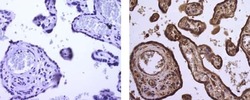
- Immunohistochemistry of formalin-fixed paraffin embedded human placenta using 20 µg/mL of Mouse IgG1 K Isotype Control Purified (left) or 20 µg/mL of Anti-Human Angiogenin Purified (right), followed by Anti-Mouse IgG Biotin, Streptavidin HRP and DAB visualization.Nuclei are counterstained with hematoxylin.

Supportive validation
- Submitted by
- Invitrogen Antibodies (provider)
- Main image

- Experimental details
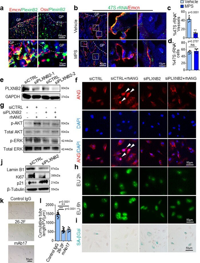
- Fig. 7 The ANG/PLXNB2 axis is essential to maintain the proliferative activity of vascular endothelial cells and protect them from senescence. a-d Three-week-old C57BL/6J mice were treated with MPS at 10 mg/m 2 /day or vehicle by daily intraperitoneal injection for 2 weeks. Double immunofluorescence staining of femoral sections using antibodies against PLXNB2 (green) and Emcn (red) or Osx (red) ( a ). Simultaneous fluorescence immunostaining of Emcn (red) and FISH analysis of 47S rRNA transcription (green) in bone tissue sections were shown in ( b ). DAPI stains nuclei blue. Boxed areas are shown at a higher magnification in corresponding panels to the right. Quantitative analysis of percentage of Emcn + blood vessels in ( c ) and non-blood vessel cells ( d ) that show positive signal of 47S rRNA transcription in primary spongiosa. Blood vessels with more than 20 green dots are considered as positive. Non-blood vessel cell with more than five green dots in nucleus are considered positive. e Immunoblot of PLXNB2 in HUVECs transfected with two individual PLXNB2 siRNAs (siPLXNB2-1, -2) or scrambled control siRNA (siCTRL). f-i HUVECs were transfected with siCTRL or siPLXNB2 for 36 h and then stimulated with 200 ng/mL rhANG or vehicle for another 12 h. Cells were fixed and immunofluorescence staining was performed using antibody against ANG (red) in ( f ). Arrow heads indicate nuclear translocation of ANG. DAPI stains nuclei blue. Immunoblot was performed using antibodies against