 Explore
 Validate
 Learn
Learn Western blot
 Immunohistochemistry
Antibody data
- Antibody Data
- Antigen structure
- References [1]
- Comments [0]
- Validations
- Western blot [1]
- Immunohistochemistry [1]
Submit
Validation data
Reference
Comment
Report error
- Product number
- HPA013138 - Provider product page

- Provider
- Atlas Antibodies
- Proper citation
- Atlas Antibodies Cat#HPA013138, RRID:AB_1855191
- Product name
- Anti-PENK
- Antibody type
- Polyclonal
- Description
- Polyclonal Antibody against Human PENK, Gene description: proenkephalin, Validated applications: IHC, WB, Uniprot ID: P01210, Storage: Store at +4°C for short term storage. Long time storage is recommended at -20°C.
- Reactivity
- Human
- Host
- Rabbit
- Conjugate
- Unconjugated
- Isotype
- IgG
- Vial size
- 100 µl
- Concentration
- 0.1 mg/ml
- Storage
- Store at +4°C for short term storage. Long time storage is recommended at -20°C.
- Handling
- The antibody solution should be gently mixed before use.
Submitted references Development and clinical validation of a seven-gene signature based on tumor stem cell-related genes to predict ovarian cancer prognosis
Wang G, Liu X, You Y, Chen S, Chang X, Yang Q
Journal of Ovarian Research 2024;17(1)
Journal of Ovarian Research 2024;17(1)
No comments: Submit comment
Enhanced validation
- Submitted by
- Atlas Antibodies (provider)
- Enhanced method
- Recombinant expression validation
- Main image
- Experimental details
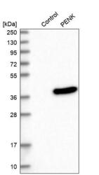
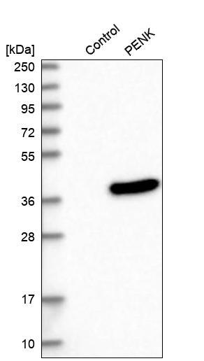
- Western blot analysis in control (vector only transfected HEK293T lysate) and PENK over-expression lysate (Co-expressed with a C-terminal myc-DDK tag (~3.1 kDa) in mammalian HEK293T cells, LY401880).
- Sample type
- Human
- Protocol
- Protocol
Supportive validation
- Submitted by
- Atlas Antibodies (provider)
- Enhanced method
- Orthogonal validation
- Main image
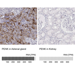
- Experimental details
- Immunohistochemistry analysis in human adrenal gland and kidney tissues using Anti-PENK antibody. Corresponding PENK RNA-seq data are presented for the same tissues.
- Sample type
- Human
- Protocol
- Protocol
