Explore
 Validate
 Learn
Learn Western blot
Antibody data
- Antibody Data
- Antigen structure
- References [2]
- Comments [0]
- Validations
- Western blot [2]
- Immunocytochemistry [2]
- Immunohistochemistry [4]
Submit
Validation data
Reference
Comment
Report error
- Product number
- GTX101786 - Provider product page

- Provider
- GeneTex
- Proper citation
- GeneTex Cat#GTX101786, RRID:AB_1240956
- Product name
- hnRNP K antibody
- Antibody type
- Polyclonal
- Reactivity
- Human, Mouse, Rat
- Host
- Rabbit
Submitted references Proteomic analysis of honokiol-induced cytotoxicity in thyroid cancer cells.
Resources for the Comprehensive Discovery of Functional RNA Elements.
Chou HC, Lu CH, Su YC, Lin LH, Yu HI, Chuang HH, Tsai YT, Liao EC, Wei YS, Yang YT, Chien YA, Yu XR, Lee YR, Chan HL
Life sciences 2018 Aug 15;207:184-204
Life sciences 2018 Aug 15;207:184-204
Resources for the Comprehensive Discovery of Functional RNA Elements.
Sundararaman B, Zhan L, Blue SM, Stanton R, Elkins K, Olson S, Wei X, Van Nostrand EL, Pratt GA, Huelga SC, Smalec BM, Wang X, Hong EL, Davidson JM, Lécuyer E, Graveley BR, Yeo GW
Molecular cell 2016 Mar 17;61(6):903-13
Molecular cell 2016 Mar 17;61(6):903-13
No comments: Submit comment
Supportive validation
- Submitted by
- GeneTex (provider)
- Main image

- Experimental details
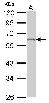
- Sample (50 ?g of whole cell lysate) A: Mouse brain 10% SDS PAGE GTX101786 diluted at 1:1000 The HRP-conjugated anti-rabbit IgG antibody (GTX213110-01) was used to detect the primary antibody.
- Submitted by
- GeneTex (provider)
- Main image
- Experimental details
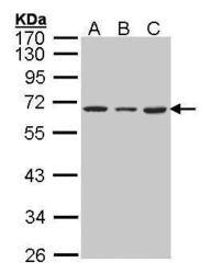
- Sample (30 ?g of whole cell lysate) A: 293T B: A431 (GTX27909) C: H1299 10% SDS PAGE GTX101786 diluted at 1:1000 The HRP-conjugated anti-rabbit IgG antibody (GTX213110-01) was used to detect the primary antibody.

Supportive validation
- Submitted by
- GeneTex (provider)
- Main image
- Experimental details
- Immunofluorescence analysis of paraformaldehyde-fixed A431, using hnRNP K(GTX101786) antibody at 1:200 dilution.
- Submitted by
- GeneTex (provider)
- Main image

- Experimental details
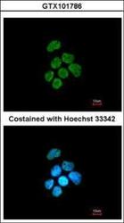
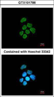
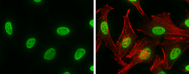
- hnRNP K antibody detects hnRNP K protein at nucleus by immunofluorescent analysis.Sample: HeLa cells were fixed in 4% paraformaldehyde at RT for 15 min.Green: hnRNP K stained by hnRNP K antibody (GTX101786) diluted at 1:500.Red: phalloidin, a cytoskeleton marker, diluted at 1:100.
Supportive validation
- Submitted by
- GeneTex (provider)
- Main image
- Experimental details
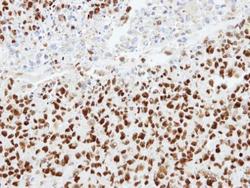
- Immunohistochemical analysis of paraffin-embedded H1299 xenograft, using hnRNP K(GTX101786) antibody at 1:100 dilution.

- Submitted by
- GeneTex (provider)
- Main image
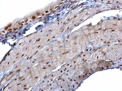
- Experimental details
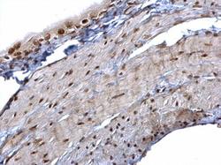
- hnRNP K antibody detects hnRNP K protein at nucleus on mouse urinary bladder by immunohistochemical analysis. Sample: Paraffin-embedded mouse urinary bladder. hnRNP K antibody (GTX101786) dilution: 1:500.
- Submitted by
- GeneTex (provider)
- Main image
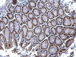
- Experimental details
- hnRNP K antibody detects hnRNP K protein at nucleus on mouse colon by immunohistochemical analysis. Sample: Paraffin-embedded mouse colon. hnRNP K antibody (GTX101786) dilution: 1:500.

- Submitted by
- GeneTex (provider)
- Main image
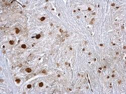
- Experimental details
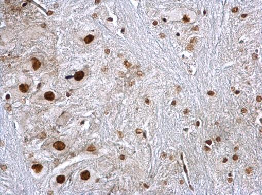
- hnRNP K antibody detects hnRNP K protein at nucleus on rat brain stem by immunohistochemical analysis. Sample: Paraffin-embedded rat brain stem. hnRNP K antibody (GTX101786) dilution: 1:500.