Explore
 Validate
 Learn
Learn Western blot
Antibody data
- Antibody Data
- Antigen structure
- References [6]
- Comments [0]
- Validations
- Western blot [4]
- Immunocytochemistry [1]
- Immunoprecipitation [1]
- Immunohistochemistry [2]
Submit
Validation data
Reference
Comment
Report error
- Product number
- GTX101089 - Provider product page

- Provider
- GeneTex
- Proper citation
- GeneTex Cat#GTX101089, RRID:AB_1952544
- Product name
- VCP antibody
- Antibody type
- Polyclonal
- Reactivity
- Human, Mouse, Rat
- Host
- Rabbit
Submitted references PTPN12/PTP-PEST Regulates Phosphorylation-Dependent Ubiquitination and Stability of Focal Adhesion Substrates in Invasive Glioblastoma Cells.
Endoplasmic Reticulum Protein TXNDC5 Augments Myocardial Fibrosis by Facilitating Extracellular Matrix Protein Folding and Redox-Sensitive Cardiac Fibroblast Activation.
Establishment of a novel three-dimensional primary culture model for hippocampal neurogenesis.
RFWD3-Mediated Ubiquitination Promotes Timely Removal of Both RPA and RAD51 from DNA Damage Sites to Facilitate Homologous Recombination.
Characterization of SET/I2PP2A isoforms in dogs.
Proteomics of muscle chronological ageing in post-menopausal women.
Chen Z, Morales JE, Guerrero PA, Sun H, McCarty JH
Cancer research 2018 Jul 15;78(14):3809-3822
Cancer research 2018 Jul 15;78(14):3809-3822
Endoplasmic Reticulum Protein TXNDC5 Augments Myocardial Fibrosis by Facilitating Extracellular Matrix Protein Folding and Redox-Sensitive Cardiac Fibroblast Activation.
Shih YC, Chen CL, Zhang Y, Mellor RL, Kanter EM, Fang Y, Wang HC, Hung CT, Nong JY, Chen HJ, Lee TH, Tseng YS, Chen CN, Wu CC, Lin SL, Yamada KA, Nerbonne JM, Yang KC
Circulation research 2018 Apr 13;122(8):1052-1068
Circulation research 2018 Apr 13;122(8):1052-1068
Establishment of a novel three-dimensional primary culture model for hippocampal neurogenesis.
Usui T, Sakurai M, Kawasaki H, Ohama T, Yamawaki H, Sato K
Physiological reports 2017 Jun;5(12)
Physiological reports 2017 Jun;5(12)
RFWD3-Mediated Ubiquitination Promotes Timely Removal of Both RPA and RAD51 from DNA Damage Sites to Facilitate Homologous Recombination.
Inano S, Sato K, Katsuki Y, Kobayashi W, Tanaka H, Nakajima K, Nakada S, Miyoshi H, Knies K, Takaori-Kondo A, Schindler D, Ishiai M, Kurumizaka H, Takata M
Molecular cell 2017 Jun 1;66(5):622-634.e8
Molecular cell 2017 Jun 1;66(5):622-634.e8
Characterization of SET/I2PP2A isoforms in dogs.
Yabe R, Fujiwara N, Mizuno T, Usui T, Ohama T, Sato K
The Journal of veterinary medical science 2014 Sep;76(9):1235-40
The Journal of veterinary medical science 2014 Sep;76(9):1235-40
Proteomics of muscle chronological ageing in post-menopausal women.
Gueugneau M, Coudy-Gandilhon C, Gourbeyre O, Chambon C, Combaret L, Polge C, Taillandier D, Attaix D, Friguet B, Maier AB, Butler-Browne G, Béchet D
BMC genomics 2014 Dec 23;15:1165
BMC genomics 2014 Dec 23;15:1165
No comments: Submit comment
Supportive validation
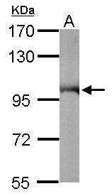
- Submitted by
- GeneTex (provider)
- Main image

- Experimental details
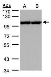
- Sample (50 ?g of whole cell lysate) A: Mouse brain 7.5% SDS PAGE GTX101089 diluted at 1:10000 The HRP-conjugated anti-rabbit IgG antibody (GTX213110-01) was used to detect the primary antibody.
- Submitted by
- GeneTex (provider)
- Main image
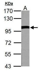
- Experimental details
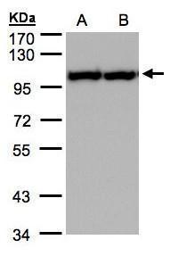
- VCP antibody detects VCP protein by western blot analysis.A. 50 ?g Rat brain lysate/extract7.5% SDS-PAGEVCP antibody (GTX101089) dilution: 1:10000 The HRP-conjugated anti-rabbit IgG antibody (GTX213110-01) was used to detect the primary antibody.
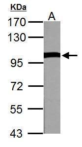
- Submitted by
- GeneTex (provider)
- Main image
- Experimental details
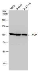
- Sample(30 ?g of whole cell lysate)A:A431(GTX27909)B:H12997.5% SDS PAGEGTX101089 diluted at 1:2000The HRP-conjugated anti-rabbit IgG antibody (GTX213110-01) was used to detect the primary antibody.
- Submitted by
- GeneTex (provider)
- Main image
- Experimental details
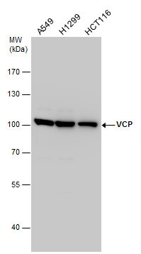
- VCP antibody detects VCP protein by western blot analysis. Various whole cell extracts (30 ?g) were separated by 7.5% SDS-PAGE, and the membrane was blotted with VCP antibody (GTX101089) diluted by 1:2000. The HRP-conjugated anti-rabbit IgG antibody (GTX213110-01) was used to detect the primary antibody.
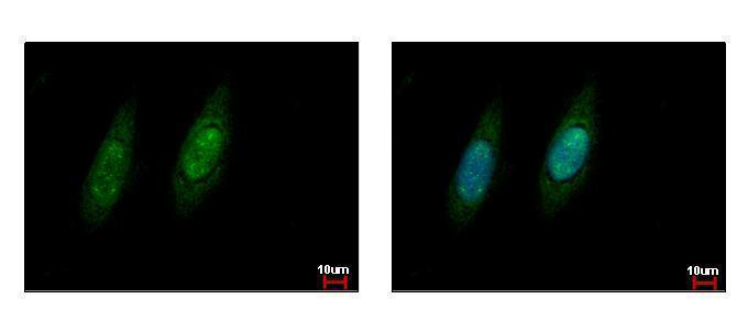
Supportive validation
- Submitted by
- GeneTex (provider)
- Main image
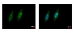
- Experimental details
- VCP antibody detects VCP protein at cytoplasm and nucleus by immunofluorescent analysis. Sample: HeLa cells were fixed in ice-cold MeOH for 5 min.Green: VCP protein stained by VCP antibody (GTX101089) diluted at 1:500.Blue: Hoechst 33343 staining.
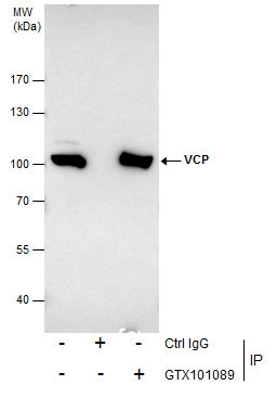
Supportive validation
- Submitted by
- GeneTex (provider)
- Main image
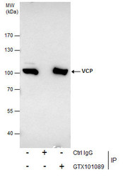
- Experimental details
- Immunoprecipitation of VCP protein from HeLa whole cell extracts using 5 £gg of VCP antibody (GTX101089).Western blot analysis was performed using VCP antibody (GTX101089).EasyBlot anti-Rabbit IgG (GTX221666-01) was used as a secondary reagent.
Supportive validation
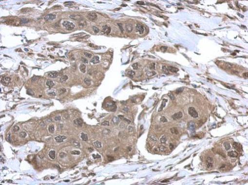
- Submitted by
- GeneTex (provider)
- Main image

- Experimental details
- Immunohistochemical analysis of paraffin-embedded human breast cancer, using VCP(GTX101089) antibody at 1:500 dilution.
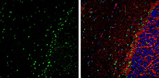
- Submitted by
- GeneTex (provider)
- Main image
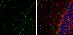
- Experimental details
- VCP antibody detects VCP Protein expression by immunohistochemical analysis.Sample: Frozen-sectioned adult mouse cerebellum. Green: VCP stained by VCP antibody (GTX101089) diluted at 1:250.Red: NF-H, stained by NF-H antibody [GT114] (GTX634289) diluted at 1:500.Blue: Fluoroshield with DAPI (GTX30920).