Explore
 Validate
 Learn
Learn Western blot
 Immunocytochemistry
Antibody data
- Antibody Data
- Antigen structure
- References [1]
- Comments [0]
- Validations
- Western blot [1]
Submit
Validation data
Reference
Comment
Report error
- Product number
- PB9454 - Provider product page

- Provider
- Boster Biological Technology
- Product name
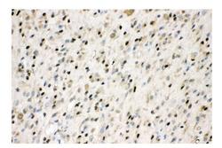
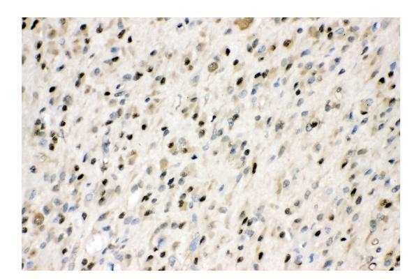
- Anti-VCP Antibody Picoband™
- Antibody type
- Polyclonal
- Description
- Polyclonal antibody for VCP detection. Host: Rabbit.Size: 100μg/vial. Tested applications: WB, IHC-P, IHC-F, ICC/IF, FCM. Reactive species: Human;Mouse;Rat. VCP information: Molecular Weight: 89322 MW; Subcellular Localization: Cytoplasm, cytosol. Endoplasmic reticulum. Nucleus. Present in the neuronal hyaline inclusion bodies specifically found in motor neurons from amyotrophic lateral sclerosis patients. Present in the Lewy bodies specifically found in neurons from Parkinson disease patients. Recruited to the cytoplasmic surface of the endoplasmic reticulum via interaction with AMFR/gp78. Following DNA double-strand breaks, recruited to the sites of damage. Recruited to stalled replication forks via interaction with SPRTN.
- Reactivity
- Human, Mouse, Rat
- Host
- Rabbit
- Vial size
- 100μg/vial
- Concentration
- Add 0.2ml of distilled water will yield a concentration of 500ug/ml.
- Storage
- At -20°C for one year. After reconstitution, at 4°C for one month. It can also be aliquoted and stored frozen at -20°C for a longer time. Avoid repeated freezing and thawing.
- Handling
- Add 0.2ml of distilled water will yield a concentration of 500ug/ml.
Submitted references KRT80 Promotes Lung Adenocarcinoma Progression and Serves as a Substrate for VCP.
Huang S, Tong W, Yang B, Ma L, Zhang J, Wang C, Xu L, Mei J
Journal of Cancer 2024;15(8):2229-2244
Journal of Cancer 2024;15(8):2229-2244
No comments: Submit comment
Supportive validation
- Submitted by
- Boster Biological Technology (provider)
- Main image
- Experimental details
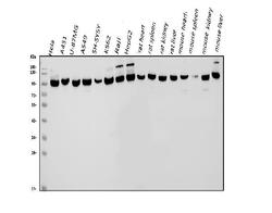
- Western blot analysis of VCP using anti-VCP antibody (PB9454). Electrophoresis was performed on a 5-20% SDS-PAGE gel at 70V (Stacking gel) / 90V (Resolving gel) for 2-3 hours. The sample well of each lane was loaded with 50ug of sample under reducing conditions. Lane 1: human Hela whole cell lysates, Lane 2: human A431 whole cell lysates, Lane 3: human U-87MG whole cell lysates, Lane 4: human A549 whole cell lysates, Lane 5: human SH-SY5Y whole cell lysates, Lane 6: human K562 whole cell lysates, Lane 7: human Raji whole cell lysates, Lane 8: human HepG2 whole cell lysates, Lane 9: rat heart tissue lysates, Lane 10: rat spleen tissue lysates, Lane 11: rat kidney tissue lysates, Lane 12: rat liver tissue lysates, Lane 13: mouse heart tissue lysates, Lane 14: mouse spleen tissue lysates, Lane 15: mouse kidney tissue lysates, Lane 16: mouse liver tissue lysates. After Electrophoresis, proteins were transferred to a Nitrocellulose membrane at 150mA for 50-90 minutes. Blocked the membrane with 5% Non-fat Milk/ TBS for 1.5 hour at RT. The membrane was incubated with rabbit anti-VCP antigen affinity purified polyclonal antibody (Catalog # PB9454) at 0.5 μg/mL overnight at 4°C, then washed with TBS-0.1%Tween 3 times with 5 minutes each and probed with a goat anti-rabbit IgG-HRP secondary antibody at a dilution of 1:10000 for 1.5 hour at RT. The signal is developed using an Enhanced Chemiluminescent detection (ECL) kit (Catalog # EK1002) with Tanon 5200 system. A specific band was detected for VCP at approximately 97KD. The expected band size for VCP is at 97KD.
- Additional image