 Explore
 Validate
 Learn
Learn Western blot
 Immunoprecipitation
Antibody data
- Antibody Data
- Antigen structure
- References [0]
- Comments [0]
- Validations
- Western blot [1]
- Immunocytochemistry [3]
- Immunohistochemistry [2]
Submit
Validation data
Reference
Comment
Report error
- Product number
- SM5062 - Provider product page

- Provider
- Acris Antibodies GmbH
- Proper citation
- Acris Antibodies GmbH Cat#SM5062, RRID:AB_1008429
- Product name
- anti TER ATPase / VCP
- Antibody type
- Monoclonal
- Antigen
- Synthetic Peptide: C G(792) G S V Y T E D N D D D L Y G (806) corresponding to amino acid residues 792-806 from Mouse VCP.
- Reactivity
- Human, Mouse, Rat
- Host
- Mouse
- Isotype
- IgG
- Antibody clone number
- 5
- Vial size
- 0.1 ml
No comments: Submit comment
Supportive validation
- Submitted by
- Acris Antibodies GmbH (provider)
- Main image

- Experimental details
- Western blot of VCP from CA46 cell lysate using SM5062.

Supportive validation
- Submitted by
- Acris Antibodies GmbH (provider)
- Main image

- Experimental details
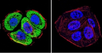
- Immunofluorescent analysis of VCP using VCP Monoclonal antibody (5) (<em>Cat.-No</em> SM5062) shows staining in ViDr colon carcinoma cells.  F-Actin staining with Phalloidin (red) and nuclei with DAPI (blue) is shown. Cells were grown on chamber slides and fixed with formaldehyde prior to staining. Cells were probed without (control) or with or an antibody recognizing VCP (<em>Cat.-No</em> SM5062) at a 1/20-1/200 overnight at 4°C, washed with PBS and incubated with a Dylight-488 conjugated secondary antibody.
- Submitted by
- Acris Antibodies GmbH (provider)
- Main image
- Experimental details
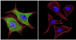
- Immunofluorescent analysis of VCP using VCP Monoclonal antibody (5) (Cat.-No SM5062) shows staining in Hela cells. F-Actin staining with Phalloidin (red) and nuclei with DAPI (blue) is shown. Cells were grown on chamber slides and fixed with formaldehyde prior to staining. Cells were probed without (control) or with or an antibody recognizing VCP (Cat.-No SM5062) at a 1/20-1/200 overnight at 4°C, washed with PBS and incubated with a Dylight-488 conjugated secondary antibody.

- Submitted by
- Acris Antibodies GmbH (provider)
- Main image

- Experimental details
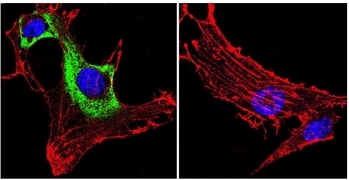
- Immunofluorescent analysis of VCP using VCP Monoclonal antibody (5) (Cat.-No SM5062) shows staining in C6 glioma cells. VCP staining (green), F-Actin staining with Phalloidin (red) and nuclei with DAPI (blue) is shown. Cells were grown on chamber slides and fixed with formaldehyde prior to staining. Cells were probed without (control) or with or an antibody recognizing VCP (Cat.-No SM5062) at a 1/20-1/200 overnight at 4°C, washed with PBS and incubated with a Dylight-488 conjugated secondary antibody.
Supportive validation
- Submitted by
- Acris Antibodies GmbH (provider)
- Main image

- Experimental details
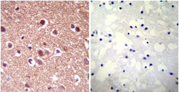
- Immunohistochemistry was performed on both normal and cancer biopsies of deparaffinized Human brain tissue tissues. To expose target proteins heat induced antigen retrieval was performed using 10mM sodium citrate (pH6.0) buffer microwaved for 8-15 minutes. Following antigen retrieval tissues were blocked in 3% BSA-PBS for 30 minutes at room temperature. Tissues were then probed at a dilution of 1/1000 with a mouse monoclonal antibody recognizing Anti-VCP (Cat.-No SM5062) or without primary antibody (negative control) overnight at 4°C in a humidified chamber. Tissues were washed extensively with PBST and endogenous peroxidase activity was quenched with a peroxidase suppressor. Detection was performed using a biotin-conjugated secondary antibody and SA-HRP followed by colorimetric detection using DAB. Tissues were counterstained with hematoxylin and prepped for mounting.
- Submitted by
- Acris Antibodies GmbH (provider)
- Main image
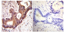
- Experimental details
- Immunohistochemistry was performed on cancer biopsies of deparaffinized Human colon carcinoma tissues. To expose target proteins, heat induced antigen retrieval was performed using 10mM sodium citrate (pH6.0) buffer, microwaved for 8-15 minutes. Following antigen retrieval tissues were blocked in 3% BSA-PBS for 30 minutes at room temperature. Tissues were then probed at a dilution of 1/1000 with SM5062 or with primary antibody (Negative Control) overnight at 4°C. in humidified chamber. Tissues were washed extensively with PBST and endogenous peroxidase activity was quenched with a peroxidase suppressor. Detection was performed using a biotin-conjugated secondary antibody and SA-HRP followed by colorimetric detection using DAB. Tissues were counterstained with hematoxylin and prepped for mounting.
