 Explore
 Validate
 Learn
Learn Western blot
 Immunocytochemistry
Antibody data
- Antibody Data
- Antigen structure
- References [0]
- Comments [0]
- Validations
- Western blot [1]
Submit
Validation data
Reference
Comment
Report error
- Product number
- M00610 - Provider product page

- Provider
- Boster Biological Technology
- Product name
- Anti-VCP Rabbit Monoclonal Antibody
- Antibody type
- Monoclonal
- Description
- Monoclonal antibody for VCP detection. Host: Rabbit.Size: 100ug/vial. Tested applications: Flow Cytometry, IP, IF, IHC, ICC, WB. Reactive species: Human, Mouse, Rat VCP information: Molecular Weight: 89322 MW; Subcellular Localization: Cytoplasm, cytosol. Endoplasmic reticulum. Nucleus. Present in the neuronal hyaline inclusion bodies specifically found in motor neurons from amyotrophic lateral sclerosis patients. Present in the Lewy bodies specifically found in neurons from Parkinson disease patients. Recruited to the cytoplasmic surface of the endoplasmic reticulum via interaction with AMFR/gp78. Following DNA double-strand breaks, recruited to the sites of damage. Recruited to stalled replication forks via interaction with SPRTN.
- Reactivity
- Human, Mouse, Rat
- Host
- Rabbit
- Antibody clone number
- AAGD-22
- Vial size
- 100ug/vial
- Concentration
- 0.5-1mg/ml, actual concentration vary by lot. Use suggested dilution ratio to decide dilution procedure.
- Storage
- At -20°C for one year. Avoid repeated freezing and thawing.
No comments: Submit comment
Supportive validation
- Submitted by
- Boster Biological Technology (provider)
- Main image
- Experimental details
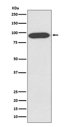
- Western blot analysis of VCP expression in HeLa cell lysate (M00610). Electrophoresis was performed on a 5-20% SDS-PAGE gel at 70V (Stacking gel) / 90V (Resolving gel) for 2-3 hours. The sample well of each lane was loaded with 50ug of sample under reducing conditions. After Electrophoresis, proteins were transferred to a Nitrocellulose membrane at 150mA for 50-90 minutes. Blocked the membrane with 5% Non-fat Milk/ TBS for 1.5 hour at RT. The membrane was incubated with rabbit anti-VCP monoclonal antibody (Catalog # M00610) overnight at 4°C, then washed with TBS-0.1%Tween 3 times with 5 minutes each and probed with a goat anti-rabbit IgG-HRP secondary antibody at a dilution of 1:10000 for 1.5 hour at RT. The signal is developed using an Enhanced Chemiluminescent detection (ECL) kit (Catalog # EK1002) with Tanon 5200 system. A specific band was detected for VCP
- Additional image


