Explore
 Validate
 Learn
LearnPA5-18290
antibody from Invitrogen Antibodies
Targeting: DLC1
ARHGAP7, DLC-1, HP, p122-RhoGAP, STARD12
 Immunocytochemistry
Antibody data
- Antibody Data
- Antigen structure
- References [1]
- Comments [0]
- Validations
- Immunocytochemistry [2]
- Immunohistochemistry [2]
- Other assay [1]
Submit
Validation data
Reference
Comment
Report error
- Product number
- PA5-18290 - Provider product page

- Provider
- Invitrogen Antibodies
- Product name
- DLC1 Polyclonal Antibody
- Antibody type
- Polyclonal
- Antigen
- Synthetic peptide
- Description
- This antibody is predicted to react with mouse and porcine based on sequence homology. This antibody is tested in Peptide ELISA: antibody detection limit dilution 64,000.
- Reactivity
- Human, Mouse
- Host
- Goat
- Isotype
- IgG
- Vial size
- 100 μg
- Concentration
- 0.5 mg/mL
- Storage
- -20°C, Avoid Freeze/Thaw Cycles
Submitted references Functional antagonism between CagA and DLC1 in gastric cancer.
Hinsenkamp I, Köhler JP, Flächsenhaar C, Hitkova I, Meessen SE, Gaiser T, Wieland T, Weiss C, Röcken C, Mowat M, Quante M, Taxauer K, Mejias-Luque R, Gerhard M, Vogelmann R, Meindl-Beinker N, Ebert M, Burgermeister E
Cell death discovery 2022 Aug 13;8(1):358
Cell death discovery 2022 Aug 13;8(1):358
No comments: Submit comment
Supportive validation
- Submitted by
- Invitrogen Antibodies (provider)
- Main image
- Experimental details
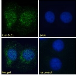
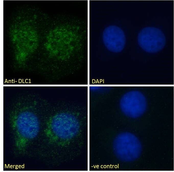
- Immunocytochemistry analysis of DLC1 using DLC1 Polyclonal Antibody (Product # PA5-18290) in paraformaldehyde fixed U251 cells, permeabilized with 0.15% Triton. Primary incubation 1hr (10 µg/mL) followed by Alexa Fluor 488 secondary antibody (2 µg/mL), showing nuclear and vesicle staining. The nuclear stain is DAPI (blue). Negative control: Unimmunized goat IgG (10 µg/mL) followed by Alexa Fluor 488 secondary antibody (2 µg/mL).
- Submitted by
- Invitrogen Antibodies (provider)
- Main image
- Experimental details
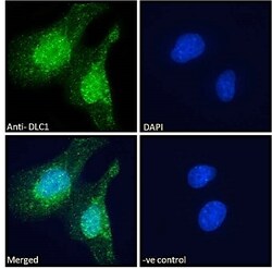
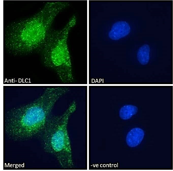
- Immunocytochemistry analysis of DLC1 using DLC1 Polyclonal Antibody (Product # PA5-18290) in paraformaldehyde fixed U2OS cells, permeabilized with 0.15% Triton. Primary incubation 1hr (10 µg/mL) followed by Alexa Fluor 488 secondary antibody (2 µg/mL), showing nuclear and cytoplasmic staining. The nuclear stain is DAPI (blue). Negative control: Unimmunized goat IgG (10 µg/mL) followed by Alexa Fluor 488 secondary antibody (2 µg/mL).
Supportive validation
- Submitted by
- Invitrogen Antibodies (provider)
- Main image

- Experimental details
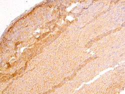
- Negative Control for DLC1 Polyclonal Antibody (Product # PA5-18290), showing immunohistochemical staining of paraffin embedded Mouse Adrenal Gland, with no primary antibody.
- Submitted by
- Invitrogen Antibodies (provider)
- Main image
- Experimental details
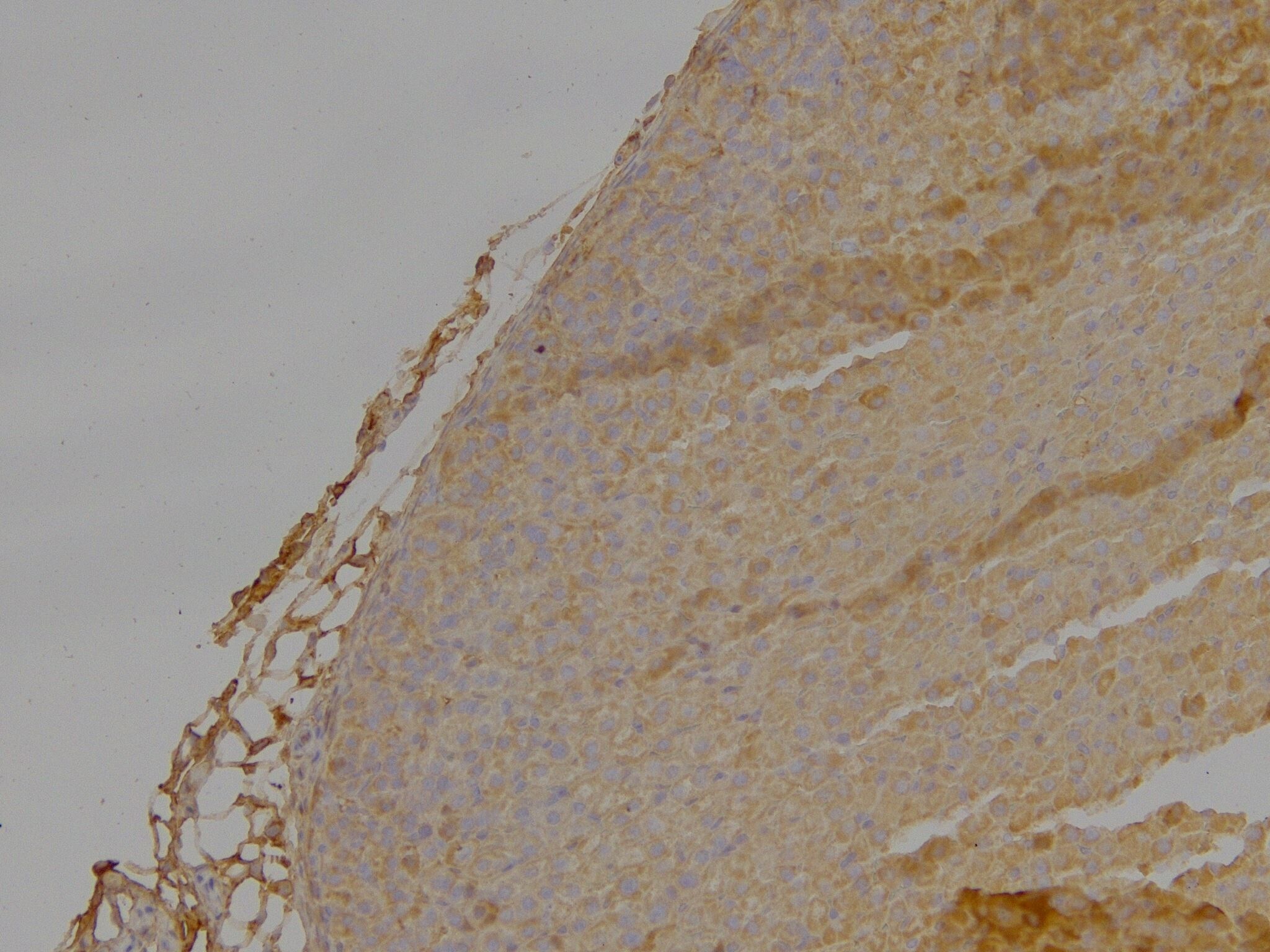
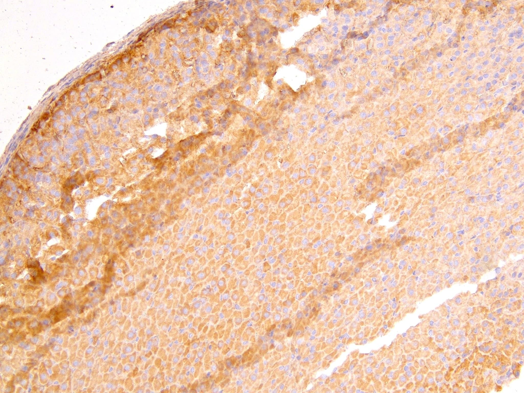
- Immunohistochemistry (PFA fixed) analysis of DLC1 using DLC1 Polyclonal Antibody (Product # PA5-18290) (4 µg/mL) in staining of paraffin embedded Mouse Adrenal Gland. Heat induced antigen retrieval with citrate buffer pH 6, HRP-staining.
Supportive validation
- Submitted by
- Invitrogen Antibodies (provider)
- Main image
- Experimental details
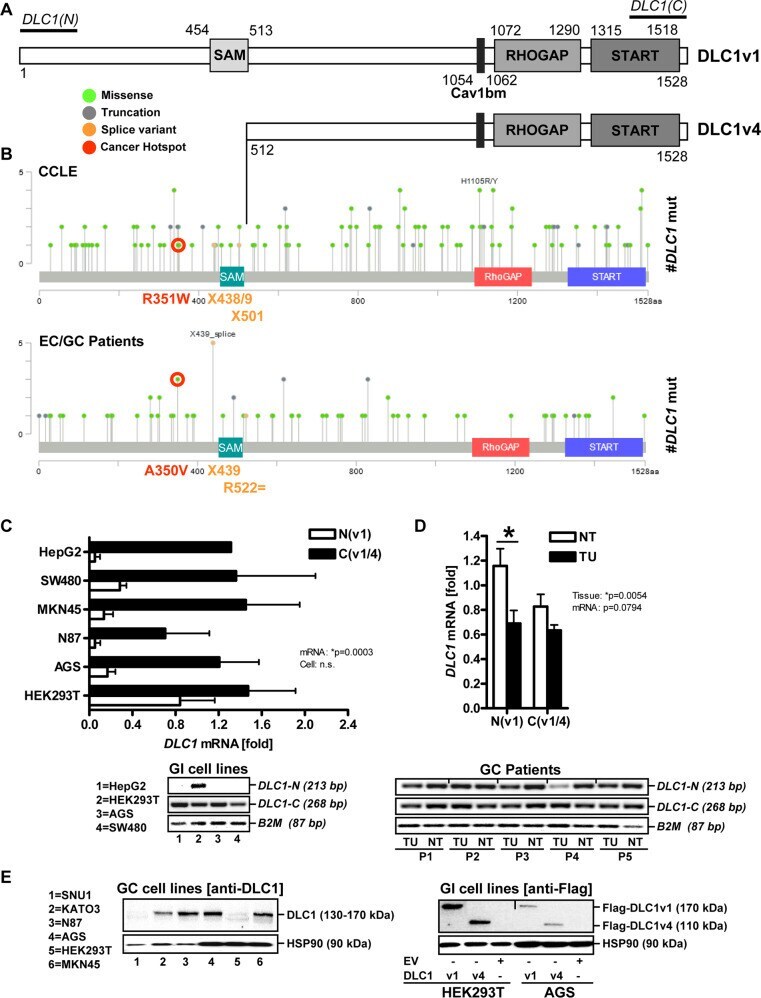
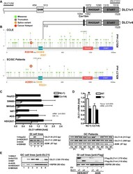
- Expression of DLC1 mRNA/protein variants in human GC cells. A Scheme of human DLC1 mRNA/protein variants. FL DLC1v1 and the N-terminally truncated DLC1v4 isoform exhibit identical C-terminal multi-domain organisation: sterile-alpha motif (SAM), serine-rich region, nuclear localisation sequence (NLS), caveolin-1 (CAV1)-binding motif (Cavbm), RHO GTPase-activating protein (GAP), and steroidogenic acute regulatory protein-related lipid-transfer (START) domains. DLC1v4 (start codon: MKLEI, aa 513-1528) used in the present study lacks the SAM region compared to DLC1v1 (aa 1-1528). B Comparison of putative driver splice mutations in the human FL DLC1v1 gene located N- and C-terminal of the SAM domain where the DLC1v4 transcript starts. Point mutation data were retrieved from cBioPortal (Table S 6 ) based on COSMIC/GISTIC entries. Cancer hotspots (red) and splice variants (yellow) are similar in human cancer cell lines (CCLE) and pan-cancer studies (from patients with EC/GC). C Detection of DLC1 mRNA variants. Total RNA from human gastrointestinal cancer and non-cancer (HEK293T) cell lines were subjected to RT-qPCR using primers directed against N- and C-terminal regions of the FL DLC1v1 cDNA. Quantitative analyses (top) and representative images from PCR-amplification products upon ethidium bromide-stained agarose gel electrophoresis (bottom). Ct-values were normalised to B2M and calculated as -fold +- S.E. (* p < 0.05 N vs. C, 2way-ANOVA with Bonferroni post-tests, n = 3 per cell