Explore
 Validate
 Learn
Learn Western blot
 Immunohistochemistry
Antibody data
- Antibody Data
- Antigen structure
- References [2]
- Comments [0]
- Validations
- Immunohistochemistry [1]
Submit
Validation data
Reference
Comment
Report error
- Product number
- AF1899 - Provider product page

- Provider
- Novus Biologicals
- Product name
- Goat Polyclonal FGFR5/FGFRL1 Antibody
- Antibody type
- Polyclonal
- Description
- Immunogen affinity purified. Detects mouse FGF R5 beta in direct ELISAs and Western blots. In Western blots, less than 5% cross-reactivity with recombinant human (rh) FGF R1 alpha (IIIb), recombinant mouse (rm) FGF R2 beta (IIIc), rmFGF R3 alpha (IIIb), rhFGF R3 alpha (IIIc), rhFGF R1 beta (IIIc), rmFGF R2 beta (IIIb), rhFGF R1 alpha (IIIc), rmFGF R2 alpha (IIIc), rhFGF R1 beta (IIIb), rmFGF R2 alpha (IIIb), rmFGF R2, and rmFGF R4 is observed.
- Reactivity
- Mouse
- Host
- Goat
- Isotype
- IgG
- Vial size
- 100 ug
- Concentration
- LYOPH
- Storage
- Use a manual defrost freezer and avoid repeated freeze-thaw cycles. 12 months from date of receipt, -20 to -70 degreesC as supplied. 1 month, 2 to 8 degreesC under sterile conditions after reconstitution. 6 months, -20 to -70 degreesC under sterile conditions after reconstitution.
Submitted references Targeted disruption of the intracellular domain of receptor FgfrL1 in mice.
The murine Fgfrl1 receptor is essential for the development of the metanephric kidney.
Bluteau G, Zhuang L, Amann R, Trueb B
PloS one 2014;9(8):e105210
PloS one 2014;9(8):e105210
The murine Fgfrl1 receptor is essential for the development of the metanephric kidney.
Gerber SD, Steinberg F, Beyeler M, Villiger PM, Trueb B
Developmental biology 2009 Nov 1;335(1):106-19
Developmental biology 2009 Nov 1;335(1):106-19
No comments: Submit comment
Supportive validation
- Submitted by
- Novus Biologicals (provider)
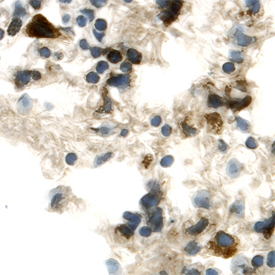
- Main image

- Experimental details
- FGF R5/FGFRL1 in Mouse Lung. FGF R5/FGFRL1 was detected in perfusion fixed frozen sections of mouse lung using Goat Anti-Mouse FGF R5/FGFRL1 Antigen Affinity-purified Polyclonal Antibody (Catalog # AF1899) at 15 µg/mL overnight at 4 °C. Tissue was stained using the Anti-Goat HRP-DAB Cell & Tissue Staining Kit (brown; Catalog # CTS008) and counterstained with hematoxylin (blue). Specific labeling was localized to the plasma membrane and cytoplasm of alveolar cells. View our protocol for Chromogenic IHC Staining of Frozen Tissue Sections.