Explore
 Validate
 Learn
Learn Immunocytochemistry
Antibody data
- Antibody Data
- Antigen structure
- References [1]
- Comments [0]
- Validations
- Immunocytochemistry [1]
- Immunohistochemistry [1]
Submit
Validation data
Reference
Comment
Report error
- Product number
- HPA036493 - Provider product page

- Provider
- Atlas Antibodies
- Proper citation
- Atlas Antibodies Cat#HPA036493, RRID:AB_10673353
- Product name
- Anti-SLC9A3
- Antibody type
- Polyclonal
- Description
- Polyclonal Antibody against Human SLC9A3, Gene description: solute carrier family 9, subfamily A (NHE3, cation proton antiporter 3), member 3, Alternative Gene Names: NHE3, Validated applications: ICC, IHC, Uniprot ID: P48764, Storage: Store at +4°C for short term storage. Long time storage is recommended at -20°C.
- Reactivity
- Human
- Host
- Rabbit
- Conjugate
- Unconjugated
- Isotype
- IgG
- Vial size
- 100 µl
- Concentration
- 0.1 mg/ml
- Storage
- Store at +4°C for short term storage. Long time storage is recommended at -20°C.
- Handling
- The antibody solution should be gently mixed before use.
Submitted references A human stomach cell type transcriptome atlas.
Öling S, Struck E, Noreen-Thorsen M, Zwahlen M, von Feilitzen K, Odeberg J, Pontén F, Lindskog C, Uhlén M, Dusart P, Butler LM
BMC biology 2024 Feb 14;22(1):36
BMC biology 2024 Feb 14;22(1):36
No comments: Submit comment
Supportive validation
- Submitted by
- Atlas Antibodies (provider)
- Main image
- Experimental details
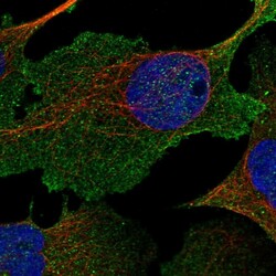
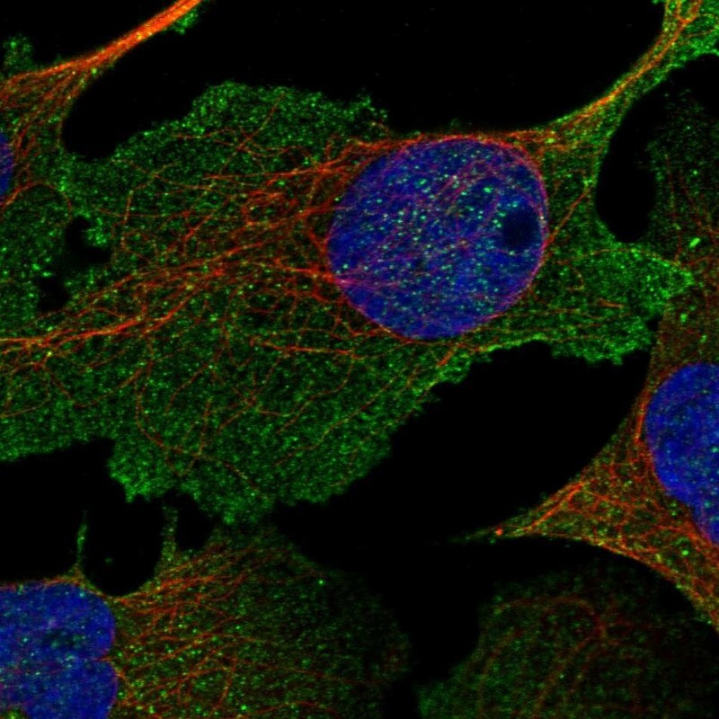
- Immunofluorescent staining of human cell line U-2 OS shows localization to plasma membrane.
- Sample type
- Human
Supportive validation
- Submitted by
- Atlas Antibodies (provider)
- Enhanced method
- Orthogonal validation
- Main image

- Experimental details
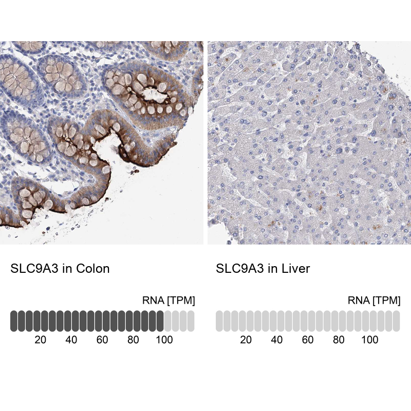
- Immunohistochemistry analysis in human colon and liver tissues using HPA036493 antibody. Corresponding SLC9A3 RNA-seq data are presented for the same tissues.
- Sample type
- Human
- Protocol
- Protocol