Explore
 Validate
 Learn
Learn Immunocytochemistry
 Immunohistochemistry
Antibody data
- Antibody Data
- Antigen structure
- References [2]
- Comments [0]
- Validations
- Immunocytochemistry [1]
Submit
Validation data
Reference
Comment
Report error
- Product number
- HPA040035 - Provider product page

- Provider
- Atlas Antibodies
- Proper citation
- Atlas Antibodies Cat#HPA040035, RRID:AB_10794207
- Product name
- Anti-MPHOSPH8
- Antibody type
- Polyclonal
- Description
- Polyclonal Antibody against Human MPHOSPH8, Gene description: M-phase phosphoprotein 8, Alternative Gene Names: HSMPP8, mpp8, Validated applications: ICC, IHC, Uniprot ID: Q99549, Storage: Store at +4°C for short term storage. Long time storage is recommended at -20°C.
- Reactivity
- Human
- Host
- Rabbit
- Conjugate
- Unconjugated
- Isotype
- IgG
- Vial size
- 100 µl
- Concentration
- 0.7 mg/ml
- Storage
- Store at +4°C for short term storage. Long time storage is recommended at -20°C.
- Handling
- The antibody solution should be gently mixed before use.
Submitted references HUSH-mediated HIV silencing is independent of TASOR phosphorylation on threonine 819
Binding to DCAF1 distinguishes TASOR and SAMHD1 degradation by HIV-2 Vpx
Vauthier V, Lasserre A, Morel M, Versapuech M, Berlioz-Torrent C, Zamborlini A, Margottin-Goguet F, Matkovic R
Retrovirology 2022;19(1)
Retrovirology 2022;19(1)
Binding to DCAF1 distinguishes TASOR and SAMHD1 degradation by HIV-2 Vpx
Johnson W, Martin M, Matkovic R, Larrous P, Morel M, Lasserre A, Vauthier V, Margottin-Goguet F
PLOS Pathogens 2021;17(10):e1009609
PLOS Pathogens 2021;17(10):e1009609
No comments: Submit comment
Supportive validation
- Submitted by
- Atlas Antibodies (provider)
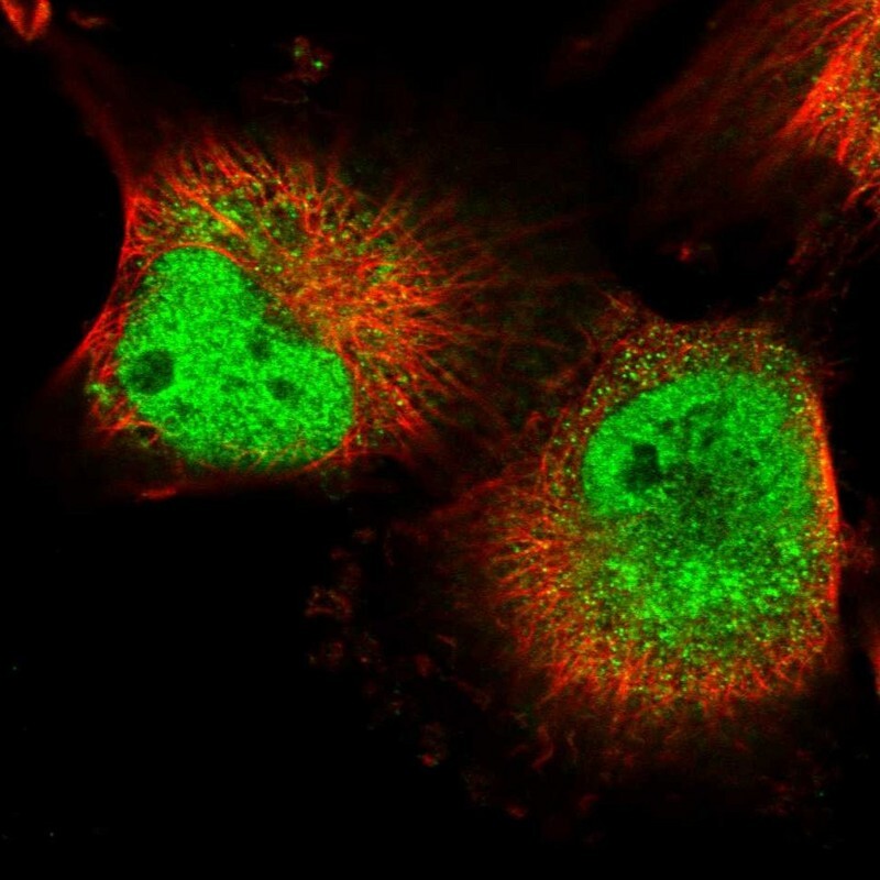
- Main image

- Experimental details
- Immunofluorescent staining of human cell line U-251 MG shows localization to nucleoplasm & cytosol.
- Sample type
- Human