Explore
 Validate
 Learn
Learn Western blot
 Immunohistochemistry
Antibody data
- Antibody Data
- Antigen structure
- References [3]
- Comments [0]
- Validations
- Western blot [1]
Submit
Validation data
Reference
Comment
Report error
- Product number
- AF2429 - Provider product page

- Provider
- R&D Systems
- Product name
- Mouse CDO Antibody
- Antibody type
- Polyclonal
- Description
- Immunogen affinity purified. Detects mouse CDO in direct ELISAs and Western blots.
- Reactivity
- Mouse
- Host
- Goat
- Conjugate
- Unconjugated
- Antigen sequence
AAC43031- Isotype
- IgG
- Vial size
- 100 ug
- Concentration
- LYOPH
- Storage
- Use a manual defrost freezer and avoid repeated freeze-thaw cycles. 12 months from date of receipt, -20 to -70 °C as supplied. 1 month, 2 to 8 °C under sterile conditions after reconstitution. 6 months, -20 to -70 °C under sterile conditions after reconstitution.
Submitted references Dehydrocorydaline promotes myogenic differentiation via p38 MAPK activation.
Syntaxin 4 regulates the surface localization of a promyogenic receptor Cdo thereby promoting myogenic differentiation.
Segregation of ipsilateral retinal ganglion cell axons at the optic chiasm requires the Shh receptor Boc.
Yoo M, Lee SJ, Kim YK, Seo DW, Baek NI, Ryu JH, Kang JS, Bae GU
Molecular medicine reports 2016 Oct;14(4):3029-36
Molecular medicine reports 2016 Oct;14(4):3029-36
Syntaxin 4 regulates the surface localization of a promyogenic receptor Cdo thereby promoting myogenic differentiation.
Yoo M, Kim BG, Lee SJ, Jeong HJ, Park JW, Seo DW, Kim YK, Lee HY, Han JW, Kang JS, Bae GU
Skeletal muscle 2015;5:28
Skeletal muscle 2015;5:28
Segregation of ipsilateral retinal ganglion cell axons at the optic chiasm requires the Shh receptor Boc.
Fabre PJ, Shimogori T, Charron F
The Journal of neuroscience : the official journal of the Society for Neuroscience 2010 Jan 6;30(1):266-75
The Journal of neuroscience : the official journal of the Society for Neuroscience 2010 Jan 6;30(1):266-75
No comments: Submit comment
Supportive validation
- Submitted by
- R&D Systems (provider)
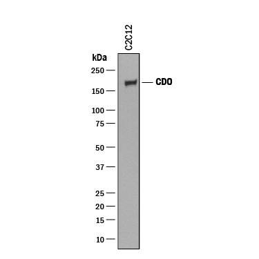
- Main image

- Experimental details
- Detection of Mouse CDO by Western Blot. Western blot shows lysate of C2C12 mouse myoblast cell line. PVDF membrane was probed with 0.25 µg/mL of Goat Anti-Mouse CDO Antigen Affinity-purified Polyclonal Antibody (Catalog # AF2429) followed by HRP-conjugated Anti-Goat IgG Secondary Antibody (Catalog # HAF019). A specific band was detected for CDO at approximately 190 kDa (as indicated). This experiment was conducted under reducing conditions and using Immunoblot Buffer Group 1.