 Explore
 Validate
 Learn
Learn Western blot
 Immunocytochemistry
Antibody data
- Antibody Data
- Antigen structure
- References [1]
- Comments [0]
- Validations
- Immunocytochemistry [1]
- Flow cytometry [1]
Submit
Validation data
Reference
Comment
Report error
- Product number
- AF4384 - Provider product page

- Provider
- R&D Systems
- Product name
- Human CDO Antibody
- Antibody type
- Polyclonal
- Description
- Antigen Affinity-purified. Detects human CDO in direct ELISAs and Western blots. In direct ELISAs and Western blots, approximately 50% cross-reactivity with recombinant mouse CDO is observed.
- Reactivity
- Human
- Host
- Sheep
- Conjugate
- Unconjugated
- Antigen sequence
NP_058648- Isotype
- IgG
- Vial size
- 100 ug
- Concentration
- LYOPH
- Storage
- Use a manual defrost freezer and avoid repeated freeze-thaw cycles. 12 months from date of receipt, -20 to -70 °C as supplied. 1 month, 2 to 8 °C under sterile conditions after reconstitution. 6 months, -20 to -70 °C under sterile conditions after reconstitution.
Submitted references Cdon acts as a Hedgehog decoy receptor during proximal-distal patterning of the optic vesicle.
Cardozo MJ, Sánchez-Arrones L, Sandonis A, Sánchez-Camacho C, Gestri G, Wilson SW, Guerrero I, Bovolenta P
Nature communications 2014 Jul 8;5:4272
Nature communications 2014 Jul 8;5:4272
No comments: Submit comment
Supportive validation
- Submitted by
- R&D Systems (provider)
- Main image
- Experimental details
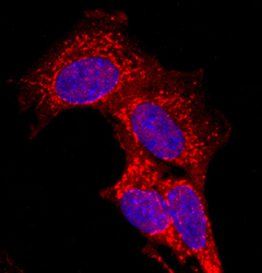
- CDO in C2C12 Mouse Cell Line. CDO was detected in immersion fixed C2C12 mouse myoblast cell line using Sheep Anti-Human CDO Antigen Affinity-purified Polyclonal Antibody (Catalog # AF4384) at 10 µg/mL for 3 hours at room temperature. Cells were stained using the NorthernLights™ 557-conjugated Anti-Sheep IgG Secondary Antibody (red; Catalog # NL010) and counterstained with DAPI (blue). Specific staining was localized to cell membranes. View our protocol for Fluorescent ICC Staining of Cells on Coverslips.

Supportive validation
- Submitted by
- R&D Systems (provider)
- Main image
- Experimental details
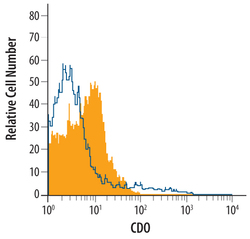
- Detection of CDO in C2C12 Mouse Cell Line by Flow Cytometry. C2C12 mouse myoblast cell line was stained with Sheep Anti-Human CDO Antigen Affinity-purified Polyclonal Antibody (Catalog # AF4384, filled histogram) or control antibody (Catalog # 5-001-A, open histogram), followed by NorthernLights™ 557-conjugated Anti-Sheep IgG Secondary Antibody (Catalog # NL010).
