Explore
 Validate
 Learn
Learn Western blot
 Immunohistochemistry
 Other assay
Antibody data
- Antibody Data
- Antigen structure
- References [57]
- Comments [0]
- Validations
- Other assay [22]
Submit
Validation data
Reference
Comment
Report error
- Product number
- 459100 - Provider product page

- Provider
- Invitrogen Antibodies
- Product name
- NDUFA9 Monoclonal Antibody (20C11B11B11)
- Antibody type
- Monoclonal
- Antigen
- Other
- Description
- Purification is near homogeneity as judged by SDS-PAGE. The antibody was produced in vitro using hybridomas grown in serum-free medium, and then purified by biochemical fractionation. 459100 detects a band at approximately 36 kDa. Use human heart mitochondria as a positive control.
- Reactivity
- Human, Mouse, Rat, Bovine
- Host
- Mouse
- Isotype
- IgG
- Antibody clone number
- 20C11B11B11
- Vial size
- 100 μL
- Concentration
- 1 mg/mL
- Storage
- 4°C
Submitted references Exercise training enhances muscle mitochondrial metabolism in diet-resistant obesity.
The Combination of Δ(9)-Tetrahydrocannabinol and Cannabidiol Suppresses Mitochondrial Respiration of Human Glioblastoma Cells via Downregulation of Specific Respiratory Chain Proteins.
Regulation of mitochondrial proteostasis by the proton gradient.
Enhancement of anaerobic glycolysis - a role of PGC-1α4 in resistance exercise.
Coding and non-coding roles of MOCCI (C15ORF48) coordinate to regulate host inflammation and immunity.
Sperm-specific COX6B2 enhances oxidative phosphorylation, proliferation, and survival in human lung adenocarcinoma.
A novel homozygous variant in MICOS13/QIL1 causes hepato-encephalopathy with mitochondrial DNA depletion syndrome.
Maintaining Myocardial Glucose Utilization in Diabetic Cardiomyopathy Accelerates Mitochondrial Dysfunction.
Leigh Syndrome Due to NDUFV1 Mutations Initially Presenting as LBSL.
Autophagy inhibition prevents glucocorticoid-increased adiposity via suppressing BAT whitening.
GLP-1 Receptor Signaling in Astrocytes Regulates Fatty Acid Oxidation, Mitochondrial Integrity, and Function.
The Mitochondria-Associated ER Membranes Are Novel Subcellular Locations Enriched for Inflammatory-Responsive MicroRNAs.
Low abundance of NDUFV2 and NDUFS4 subunits of the hydrophilic complex I domain and VDAC1 predicts mammalian longevity.
Chloramphenicol Mitigates Oxidative Stress by Inhibiting Translation of Mitochondrial Complex I in Dopaminergic Neurons of Toxin-Induced Parkinson's Disease Model.
Mitochondrial supercomplex assembly promotes breast and endometrial tumorigenesis by metabolic alterations and enhanced hypoxia tolerance.
Fascin Controls Metastatic Colonization and Mitochondrial Oxidative Phosphorylation by Remodeling Mitochondrial Actin Filaments.
Depletion of Mitochondrial DNA in Differentiated Retinal Pigment Epithelial Cells.
Mutations in the mitochondrial complex I assembly factor NDUFAF6 cause isolated bilateral striatal necrosis and progressive dystonia in childhood.
Fractionated mitochondrial magnetic separation for isolation of synaptic mitochondria from brain tissue.
Peroxynitrite supports a metabolic reprogramming in merlin-deficient Schwann cells and promotes cell survival.
Caloric Restriction Induces MicroRNAs to Improve Mitochondrial Proteostasis.
Loss of the mitochondrial i-AAA protease YME1L leads to ocular dysfunction and spinal axonopathy.
Coenzyme Q10 protects against burn-induced mitochondrial dysfunction and impaired insulin signaling in mouse skeletal muscle.
IL-15 improves skeletal muscle oxidative metabolism and glucose uptake in association with increased respiratory chain supercomplex formation and AMPK pathway activation.
The mitochondrial deoxyguanosine kinase is required for cancer cell stemness in lung adenocarcinoma.
Age-related sex differences in the expression of important disease-linked mitochondrial proteins in mice.
COX6A2 variants cause a muscle-specific cytochrome c oxidase deficiency.
Absence of TXNIP in Humans Leads to Lactic Acidosis and Low Serum Methionine Linked to Deficient Respiration on Pyruvate.
The Expression of Uncoupling Protein 3 Coincides With the Fatty Acid Oxidation Type of Metabolism in Adult Murine Heart.
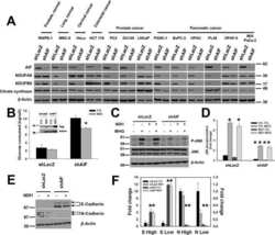
AIF promotes a JNK1-mediated cadherin switch independently of respiratory chain stabilization.
Mild Impairment of Mitochondrial OXPHOS Promotes Fatty Acid Utilization in POMC Neurons and Improves Glucose Homeostasis in Obesity.
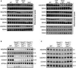
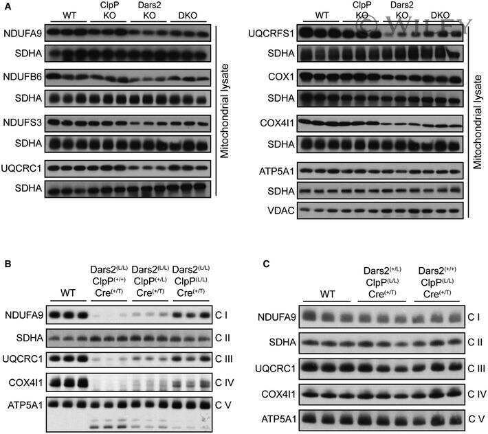
CLPP deficiency protects against metabolic syndrome but hinders adaptive thermogenesis.
Toxoplasma gondii GRA8 induces ATP5A1-SIRT3-mediated mitochondrial metabolic resuscitation: a potential therapy for sepsis.
Alternative assembly of respiratory complex II connects energy stress to metabolic checkpoints.
Mitochondrial dysfunction underlies cognitive defects as a result of neural stem cell depletion and impaired neurogenesis.
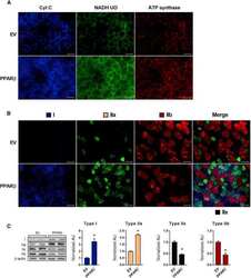
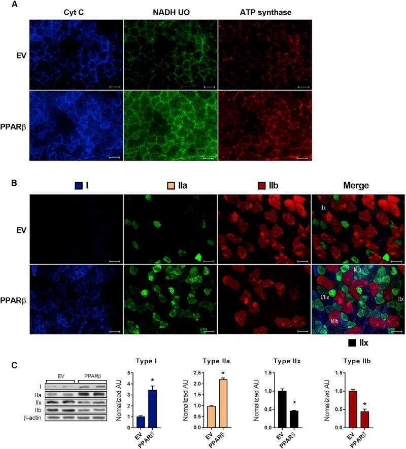
PPARβ Is Essential for Maintaining Normal Levels of PGC-1α and Mitochondria and for the Increase in Muscle Mitochondria Induced by Exercise.
(D)-Glutamate is metabolized in the heart mitochondria.
CLUH regulates mitochondrial metabolism by controlling translation and decay of target mRNAs.
Burn-induced muscle metabolic derangements and mitochondrial dysfunction are associated with activation of HIF-1α and mTORC1: Role of protein farnesylation.
Selective Disruption of Respiratory Supercomplexes as a New Strategy to Suppress Her2(high) Breast Cancer.
Twinkle overexpression prevents cardiac rupture after myocardial infarction by alleviating impaired mitochondrial biogenesis.
Amyloid β-peptides interfere with mitochondrial preprotein import competence by a coaggregation process.
Loss of CLPP alleviates mitochondrial cardiomyopathy without affecting the mammalian UPRmt.
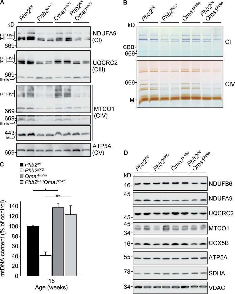
Loss of OMA1 delays neurodegeneration by preventing stress-induced OPA1 processing in mitochondria.
Basal metabolic state governs AIF-dependent growth support in pancreatic cancer cells.
Overexpression of TFAM or twinkle increases mtDNA copy number and facilitates cardioprotection associated with limited mitochondrial oxidative stress.
Cysteine dietary supplementation reverses the decrease in mitochondrial ROS production at complex I induced by methionine restriction.
Essential role of mitochondrial Ca2+ uniporter in the generation of mitochondrial pH gradient and metabolism-secretion coupling in insulin-releasing cells.
A keratin scaffold regulates epidermal barrier formation, mitochondrial lipid composition, and activity.
Imbalanced OPA1 processing and mitochondrial fragmentation cause heart failure in mice.
CNC-bZIP protein Nrf1-dependent regulation of glucose-stimulated insulin secretion.
P150glued-associated disorders are caused by activation of intrinsic apoptotic pathway.
Cytosolic p53 inhibits Parkin-mediated mitophagy and promotes mitochondrial dysfunction in the mouse heart.
β-Adrenergic stimulation does not activate p38 MAP kinase or induce PGC-1α in skeletal muscle.
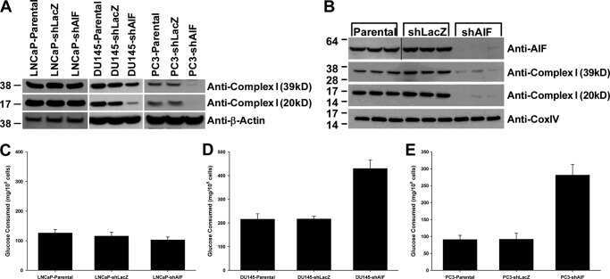
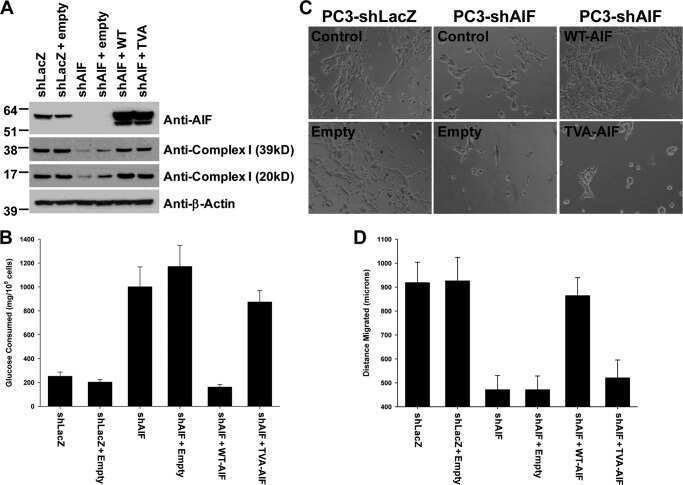
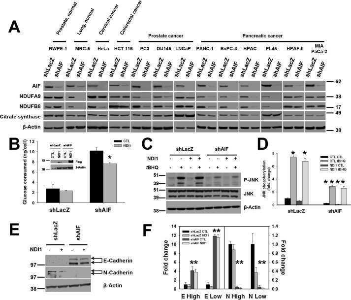
The enzymatic activity of apoptosis-inducing factor supports energy metabolism benefiting the growth and invasiveness of advanced prostate cancer cells.
Mitochondrial DNA toxicity compromises mitochondrial dynamics and induces hippocampal antioxidant defenses.
Unexpected vascular enrichment of SCO1 over SCO2 in mammalian tissues: implications for human mitochondrial disease.
Pileggi CA, Blondin DP, Hooks BG, Parmar G, Alecu I, Patten DA, Cuillerier A, O'Dwyer C, Thrush AB, Fullerton MD, Bennett SA, Doucet É, Haman F, Cuperlovic-Culf M, McPherson R, Dent RRM, Harper ME
EBioMedicine 2022 Sep;83:104192
EBioMedicine 2022 Sep;83:104192
The Combination of Δ(9)-Tetrahydrocannabinol and Cannabidiol Suppresses Mitochondrial Respiration of Human Glioblastoma Cells via Downregulation of Specific Respiratory Chain Proteins.
Rupprecht A, Theisen U, Wendt F, Frank M, Hinz B
Cancers 2022 Jun 27;14(13)
Cancers 2022 Jun 27;14(13)
Regulation of mitochondrial proteostasis by the proton gradient.
Patron M, Tarasenko D, Nolte H, Kroczek L, Ghosh M, Ohba Y, Lasarzewski Y, Ahmadi ZA, Cabrera-Orefice A, Eyiama A, Kellermann T, Rugarli EI, Brandt U, Meinecke M, Langer T
The EMBO journal 2022 Aug 16;41(16):e110476
The EMBO journal 2022 Aug 16;41(16):e110476
Enhancement of anaerobic glycolysis - a role of PGC-1α4 in resistance exercise.
Koh JH, Pataky MW, Dasari S, Klaus KA, Vuckovic I, Ruegsegger GN, Kumar AP, Robinson MM, Nair KS
Nature communications 2022 Apr 28;13(1):2324
Nature communications 2022 Apr 28;13(1):2324
Coding and non-coding roles of MOCCI (C15ORF48) coordinate to regulate host inflammation and immunity.
Lee CQE, Kerouanton B, Chothani S, Zhang S, Chen Y, Mantri CK, Hock DH, Lim R, Nadkarni R, Huynh VT, Lim D, Chew WL, Zhong FL, Stroud DA, Schafer S, Tergaonkar V, St John AL, Rackham OJL, Ho L
Nature communications 2021 Apr 9;12(1):2130
Nature communications 2021 Apr 9;12(1):2130
Sperm-specific COX6B2 enhances oxidative phosphorylation, proliferation, and survival in human lung adenocarcinoma.
Cheng CC, Wooten J, Gibbs ZA, McGlynn K, Mishra P, Whitehurst AW
eLife 2020 Sep 29;9
eLife 2020 Sep 29;9
A novel homozygous variant in MICOS13/QIL1 causes hepato-encephalopathy with mitochondrial DNA depletion syndrome.
Kishita Y, Shimura M, Kohda M, Akita M, Imai-Okazaki A, Yatsuka Y, Nakajima Y, Ito T, Ohtake A, Murayama K, Okazaki Y
Molecular genetics & genomic medicine 2020 Oct;8(10):e1427
Molecular genetics & genomic medicine 2020 Oct;8(10):e1427
Maintaining Myocardial Glucose Utilization in Diabetic Cardiomyopathy Accelerates Mitochondrial Dysfunction.
Wende AR, Schell JC, Ha CM, Pepin ME, Khalimonchuk O, Schwertz H, Pereira RO, Brahma MK, Tuinei J, Contreras-Ferrat A, Wang L, Andrizzi CA, Olsen CD, Bradley WE, Dell'Italia LJ, Dillmann WH, Litwin SE, Abel ED
Diabetes 2020 Oct;69(10):2094-2111
Diabetes 2020 Oct;69(10):2094-2111
Leigh Syndrome Due to NDUFV1 Mutations Initially Presenting as LBSL.
Borna NN, Kishita Y, Sakai N, Hamada Y, Kamagata K, Kohda M, Ohtake A, Murayama K, Okazaki Y
Genes 2020 Nov 9;11(11)
Genes 2020 Nov 9;11(11)
Autophagy inhibition prevents glucocorticoid-increased adiposity via suppressing BAT whitening.
Deng J, Guo Y, Yuan F, Chen S, Yin H, Jiang X, Jiao F, Wang F, Ji H, Hu G, Ying H, Chen Y, Zhai Q, Xiao F, Guo F
Autophagy 2020 Mar;16(3):451-465
Autophagy 2020 Mar;16(3):451-465
GLP-1 Receptor Signaling in Astrocytes Regulates Fatty Acid Oxidation, Mitochondrial Integrity, and Function.
Timper K, Del Río-Martín A, Cremer AL, Bremser S, Alber J, Giavalisco P, Varela L, Heilinger C, Nolte H, Trifunovic A, Horvath TL, Kloppenburg P, Backes H, Brüning JC
Cell metabolism 2020 Jun 2;31(6):1189-1205.e13
Cell metabolism 2020 Jun 2;31(6):1189-1205.e13
The Mitochondria-Associated ER Membranes Are Novel Subcellular Locations Enriched for Inflammatory-Responsive MicroRNAs.
Wang WX, Prajapati P, Nelson PT, Springer JE
Molecular neurobiology 2020 Jul;57(7):2996-3013
Molecular neurobiology 2020 Jul;57(7):2996-3013
Low abundance of NDUFV2 and NDUFS4 subunits of the hydrophilic complex I domain and VDAC1 predicts mammalian longevity.
Mota-Martorell N, Jove M, Pradas I, Sanchez I, Gómez J, Naudi A, Barja G, Pamplona R
Redox biology 2020 Jul;34:101539
Redox biology 2020 Jul;34:101539
Chloramphenicol Mitigates Oxidative Stress by Inhibiting Translation of Mitochondrial Complex I in Dopaminergic Neurons of Toxin-Induced Parkinson's Disease Model.
Han J, Kim SJ, Ryu MJ, Jang Y, Lee MJ, Ju X, Lee YL, Cui J, Shong M, Heo JY, Kweon GR
Oxidative medicine and cellular longevity 2019;2019:4174803
Oxidative medicine and cellular longevity 2019;2019:4174803
Mitochondrial supercomplex assembly promotes breast and endometrial tumorigenesis by metabolic alterations and enhanced hypoxia tolerance.
Ikeda K, Horie-Inoue K, Suzuki T, Hobo R, Nakasato N, Takeda S, Inoue S
Nature communications 2019 Sep 11;10(1):4108
Nature communications 2019 Sep 11;10(1):4108
Fascin Controls Metastatic Colonization and Mitochondrial Oxidative Phosphorylation by Remodeling Mitochondrial Actin Filaments.
Lin S, Huang C, Gunda V, Sun J, Chellappan SP, Li Z, Izumi V, Fang B, Koomen J, Singh PK, Hao J, Yang S
Cell reports 2019 Sep 10;28(11):2824-2836.e8
Cell reports 2019 Sep 10;28(11):2824-2836.e8
Depletion of Mitochondrial DNA in Differentiated Retinal Pigment Epithelial Cells.
Hu X, Calton MA, Tang S, Vollrath D
Scientific reports 2019 Oct 25;9(1):15355
Scientific reports 2019 Oct 25;9(1):15355
Mutations in the mitochondrial complex I assembly factor NDUFAF6 cause isolated bilateral striatal necrosis and progressive dystonia in childhood.
Baide-Mairena H, Gaudó P, Marti-Sánchez L, Emperador S, Sánchez-Montanez A, Alonso-Luengo O, Correa M, Grau AM, Ortigoza-Escobar JD, Artuch R, Vázquez E, Del Toro M, Garrido-Pérez N, Ruiz-Pesini E, Montoya J, Bayona-Bafaluy MP, Pérez-Dueñas B
Molecular genetics and metabolism 2019 Mar;126(3):250-258
Molecular genetics and metabolism 2019 Mar;126(3):250-258
Fractionated mitochondrial magnetic separation for isolation of synaptic mitochondria from brain tissue.
Hubbard WB, Harwood CL, Prajapati P, Springer JE, Saatman KE, Sullivan PG
Scientific reports 2019 Jul 4;9(1):9656
Scientific reports 2019 Jul 4;9(1):9656
Peroxynitrite supports a metabolic reprogramming in merlin-deficient Schwann cells and promotes cell survival.
Pestoni JC, Klingeman Plati S, Valdivia Camacho OD, Fuse MA, Onatunde M, Sparrow NA, Karajannis MA, Fernández-Valle C, Franco MC
The Journal of biological chemistry 2019 Jul 26;294(30):11354-11368
The Journal of biological chemistry 2019 Jul 26;294(30):11354-11368
Caloric Restriction Induces MicroRNAs to Improve Mitochondrial Proteostasis.
Zhang R, Wang X, Qu JH, Liu B, Zhang P, Zhang T, Fan PC, Wang XM, Xiao GY, Su Y, Xie Y, Liu Y, Pei JF, Zhang ZQ, Hao DL, Xu P, Chen HZ, Liu DP
iScience 2019 Jul 26;17:155-166
iScience 2019 Jul 26;17:155-166
Loss of the mitochondrial i-AAA protease YME1L leads to ocular dysfunction and spinal axonopathy.
Sprenger HG, Wani G, Hesseling A, König T, Patron M, MacVicar T, Ahola S, Wai T, Barth E, Rugarli EI, Bergami M, Langer T
EMBO molecular medicine 2019 Jan;11(1)
EMBO molecular medicine 2019 Jan;11(1)
Coenzyme Q10 protects against burn-induced mitochondrial dysfunction and impaired insulin signaling in mouse skeletal muscle.
Nakazawa H, Ikeda K, Shinozaki S, Yasuhara S, Yu YM, Martyn JAJ, Tompkins RG, Yorozu T, Inoue S, Kaneki M
FEBS open bio 2019 Feb;9(2):348-363
FEBS open bio 2019 Feb;9(2):348-363
IL-15 improves skeletal muscle oxidative metabolism and glucose uptake in association with increased respiratory chain supercomplex formation and AMPK pathway activation.
Nadeau L, Patten DA, Caron A, Garneau L, Pinault-Masson E, Foretz M, Haddad P, Anderson BG, Quinn LS, Jardine K, McBurney MW, Pistilli EE, Harper ME, Aguer C
Biochimica et biophysica acta. General subjects 2019 Feb;1863(2):395-407
Biochimica et biophysica acta. General subjects 2019 Feb;1863(2):395-407
The mitochondrial deoxyguanosine kinase is required for cancer cell stemness in lung adenocarcinoma.
Lin S, Huang C, Sun J, Bollt O, Wang X, Martine E, Kang J, Taylor MD, Fang B, Singh PK, Koomen J, Hao J, Yang S
EMBO molecular medicine 2019 Dec;11(12):e10849
EMBO molecular medicine 2019 Dec;11(12):e10849
Age-related sex differences in the expression of important disease-linked mitochondrial proteins in mice.
Moschinger M, Hilse KE, Rupprecht A, Zeitz U, Erben RG, Rülicke T, Pohl EE
Biology of sex differences 2019 Dec 5;10(1):56
Biology of sex differences 2019 Dec 5;10(1):56
COX6A2 variants cause a muscle-specific cytochrome c oxidase deficiency.
Inoue M, Uchino S, Iida A, Noguchi S, Hayashi S, Takahashi T, Fujii K, Komaki H, Takeshita E, Nonaka I, Okada Y, Yoshizawa T, Van Lommel L, Schuit F, Goto YI, Mimaki M, Nishino I
Annals of neurology 2019 Aug;86(2):193-202
Annals of neurology 2019 Aug;86(2):193-202
Absence of TXNIP in Humans Leads to Lactic Acidosis and Low Serum Methionine Linked to Deficient Respiration on Pyruvate.
Katsu-Jiménez Y, Vázquez-Calvo C, Maffezzini C, Halldin M, Peng X, Freyer C, Wredenberg A, Giménez-Cassina A, Wedell A, Arnér ESJ
Diabetes 2019 Apr;68(4):709-723
Diabetes 2019 Apr;68(4):709-723
The Expression of Uncoupling Protein 3 Coincides With the Fatty Acid Oxidation Type of Metabolism in Adult Murine Heart.
Hilse KE, Rupprecht A, Egerbacher M, Bardakji S, Zimmermann L, Wulczyn AEMS, Pohl EE
Frontiers in physiology 2018;9:747
Frontiers in physiology 2018;9:747
AIF promotes a JNK1-mediated cadherin switch independently of respiratory chain stabilization.
Scott AJ, Walker SA, Krank JJ, Wilkinson AS, Johnson KM, Lewis EM, Wilkinson JC
The Journal of biological chemistry 2018 Sep 21;293(38):14707-14722
The Journal of biological chemistry 2018 Sep 21;293(38):14707-14722
Mild Impairment of Mitochondrial OXPHOS Promotes Fatty Acid Utilization in POMC Neurons and Improves Glucose Homeostasis in Obesity.
Timper K, Paeger L, Sánchez-Lasheras C, Varela L, Jais A, Nolte H, Vogt MC, Hausen AC, Heilinger C, Evers N, Pospisilik JA, Penninger JM, Taylor EB, Horvath TL, Kloppenburg P, Brüning JC
Cell reports 2018 Oct 9;25(2):383-397.e10
Cell reports 2018 Oct 9;25(2):383-397.e10
CLPP deficiency protects against metabolic syndrome but hinders adaptive thermogenesis.
Becker C, Kukat A, Szczepanowska K, Hermans S, Senft K, Brandscheid CP, Maiti P, Trifunovic A
EMBO reports 2018 May;19(5)
EMBO reports 2018 May;19(5)
Toxoplasma gondii GRA8 induces ATP5A1-SIRT3-mediated mitochondrial metabolic resuscitation: a potential therapy for sepsis.
Kim YR, Kim JS, Yun JS, Kim S, Kim SY, Jang K, Yang CS
Experimental & molecular medicine 2018 Mar 30;50(3):e464
Experimental & molecular medicine 2018 Mar 30;50(3):e464
Alternative assembly of respiratory complex II connects energy stress to metabolic checkpoints.
Bezawork-Geleta A, Wen H, Dong L, Yan B, Vider J, Boukalova S, Krobova L, Vanova K, Zobalova R, Sobol M, Hozak P, Novais SM, Caisova V, Abaffy P, Naraine R, Pang Y, Zaw T, Zhang P, Sindelka R, Kubista M, Zuryn S, Molloy MP, Berridge MV, Pacak K, Rohlena J, Park S, Neuzil J
Nature communications 2018 Jun 7;9(1):2221
Nature communications 2018 Jun 7;9(1):2221
Mitochondrial dysfunction underlies cognitive defects as a result of neural stem cell depletion and impaired neurogenesis.
Khacho M, Clark A, Svoboda DS, MacLaurin JG, Lagace DC, Park DS, Slack RS
Human molecular genetics 2017 Sep 1;26(17):3327-3341
Human molecular genetics 2017 Sep 1;26(17):3327-3341
PPARβ Is Essential for Maintaining Normal Levels of PGC-1α and Mitochondria and for the Increase in Muscle Mitochondria Induced by Exercise.
Koh JH, Hancock CR, Terada S, Higashida K, Holloszy JO, Han DH
Cell metabolism 2017 May 2;25(5):1176-1185.e5
Cell metabolism 2017 May 2;25(5):1176-1185.e5
(D)-Glutamate is metabolized in the heart mitochondria.
Ariyoshi M, Katane M, Hamase K, Miyoshi Y, Nakane M, Hoshino A, Okawa Y, Mita Y, Kaimoto S, Uchihashi M, Fukai K, Ono K, Tateishi S, Hato D, Yamanaka R, Honda S, Fushimura Y, Iwai-Kanai E, Ishihara N, Mita M, Homma H, Matoba S
Scientific reports 2017 Mar 7;7:43911
Scientific reports 2017 Mar 7;7:43911
CLUH regulates mitochondrial metabolism by controlling translation and decay of target mRNAs.
Schatton D, Pla-Martin D, Marx MC, Hansen H, Mourier A, Nemazanyy I, Pessia A, Zentis P, Corona T, Kondylis V, Barth E, Schauss AC, Velagapudi V, Rugarli EI
The Journal of cell biology 2017 Mar 6;216(3):675-693
The Journal of cell biology 2017 Mar 6;216(3):675-693
Burn-induced muscle metabolic derangements and mitochondrial dysfunction are associated with activation of HIF-1α and mTORC1: Role of protein farnesylation.
Nakazawa H, Ikeda K, Shinozaki S, Kobayashi M, Ikegami Y, Fu M, Nakamura T, Yasuhara S, Yu YM, Martyn JAJ, Tompkins RG, Shimokado K, Yorozu T, Ito H, Inoue S, Kaneki M
Scientific reports 2017 Jul 26;7(1):6618
Scientific reports 2017 Jul 26;7(1):6618
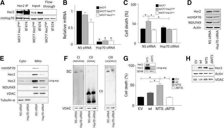
Selective Disruption of Respiratory Supercomplexes as a New Strategy to Suppress Her2(high) Breast Cancer.
Rohlenova K, Sachaphibulkij K, Stursa J, Bezawork-Geleta A, Blecha J, Endaya B, Werner L, Cerny J, Zobalova R, Goodwin J, Spacek T, Alizadeh Pesdar E, Yan B, Nguyen MN, Vondrusova M, Sobol M, Jezek P, Hozak P, Truksa J, Rohlena J, Dong LF, Neuzil J
Antioxidants & redox signaling 2017 Jan 10;26(2):84-103
Antioxidants & redox signaling 2017 Jan 10;26(2):84-103
Twinkle overexpression prevents cardiac rupture after myocardial infarction by alleviating impaired mitochondrial biogenesis.
Inoue T, Ikeda M, Ide T, Fujino T, Matsuo Y, Arai S, Saku K, Sunagawa K
American journal of physiology. Heart and circulatory physiology 2016 Sep 1;311(3):H509-19
American journal of physiology. Heart and circulatory physiology 2016 Sep 1;311(3):H509-19
Amyloid β-peptides interfere with mitochondrial preprotein import competence by a coaggregation process.
Cenini G, Rüb C, Bruderek M, Voos W
Molecular biology of the cell 2016 Nov 1;27(21):3257-3272
Molecular biology of the cell 2016 Nov 1;27(21):3257-3272
Loss of CLPP alleviates mitochondrial cardiomyopathy without affecting the mammalian UPRmt.
Seiferling D, Szczepanowska K, Becker C, Senft K, Hermans S, Maiti P, König T, Kukat A, Trifunovic A
EMBO reports 2016 Jul;17(7):953-64
EMBO reports 2016 Jul;17(7):953-64
Loss of OMA1 delays neurodegeneration by preventing stress-induced OPA1 processing in mitochondria.
Korwitz A, Merkwirth C, Richter-Dennerlein R, Tröder SE, Sprenger HG, Quirós PM, López-Otín C, Rugarli EI, Langer T
The Journal of cell biology 2016 Jan 18;212(2):157-66
The Journal of cell biology 2016 Jan 18;212(2):157-66
Basal metabolic state governs AIF-dependent growth support in pancreatic cancer cells.
Scott AJ, Wilkinson AS, Wilkinson JC
BMC cancer 2016 Apr 23;16:286
BMC cancer 2016 Apr 23;16:286
Overexpression of TFAM or twinkle increases mtDNA copy number and facilitates cardioprotection associated with limited mitochondrial oxidative stress.
Ikeda M, Ide T, Fujino T, Arai S, Saku K, Kakino T, Tyynismaa H, Yamasaki T, Yamada K, Kang D, Suomalainen A, Sunagawa K
PloS one 2015;10(3):e0119687
PloS one 2015;10(3):e0119687
Cysteine dietary supplementation reverses the decrease in mitochondrial ROS production at complex I induced by methionine restriction.
Gomez A, Gomez J, Lopez Torres M, Naudi A, Mota-Martorell N, Pamplona R, Barja G
Journal of bioenergetics and biomembranes 2015 Jun;47(3):199-208
Journal of bioenergetics and biomembranes 2015 Jun;47(3):199-208
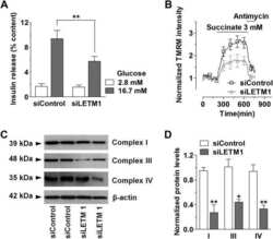
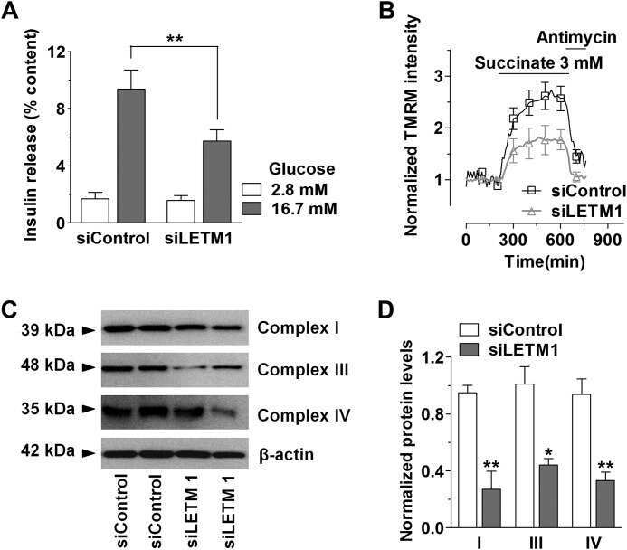
Essential role of mitochondrial Ca2+ uniporter in the generation of mitochondrial pH gradient and metabolism-secretion coupling in insulin-releasing cells.
Quan X, Nguyen TT, Choi SK, Xu S, Das R, Cha SK, Kim N, Han J, Wiederkehr A, Wollheim CB, Park KS
The Journal of biological chemistry 2015 Feb 13;290(7):4086-96
The Journal of biological chemistry 2015 Feb 13;290(7):4086-96
A keratin scaffold regulates epidermal barrier formation, mitochondrial lipid composition, and activity.
Kumar V, Bouameur JE, Bär J, Rice RH, Hornig-Do HT, Roop DR, Schwarz N, Brodesser S, Thiering S, Leube RE, Wiesner RJ, Vijayaraj P, Brazel CB, Heller S, Binder H, Löffler-Wirth H, Seibel P, Magin TM
The Journal of cell biology 2015 Dec 7;211(5):1057-75
The Journal of cell biology 2015 Dec 7;211(5):1057-75
Imbalanced OPA1 processing and mitochondrial fragmentation cause heart failure in mice.
Wai T, García-Prieto J, Baker MJ, Merkwirth C, Benit P, Rustin P, Rupérez FJ, Barbas C, Ibañez B, Langer T
Science (New York, N.Y.) 2015 Dec 4;350(6265):aad0116
Science (New York, N.Y.) 2015 Dec 4;350(6265):aad0116
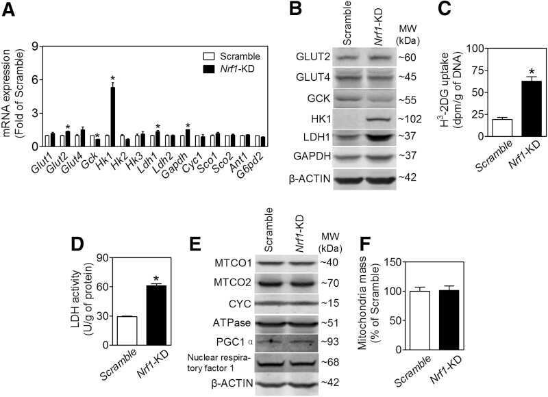
CNC-bZIP protein Nrf1-dependent regulation of glucose-stimulated insulin secretion.
Zheng H, Fu J, Xue P, Zhao R, Dong J, Liu D, Yamamoto M, Tong Q, Teng W, Qu W, Zhang Q, Andersen ME, Pi J
Antioxidants & redox signaling 2015 Apr 1;22(10):819-31
Antioxidants & redox signaling 2015 Apr 1;22(10):819-31
P150glued-associated disorders are caused by activation of intrinsic apoptotic pathway.
Ishikawa K, Saiki S, Furuya N, Yamada D, Imamichi Y, Li Y, Kawajiri S, Sasaki H, Koike M, Tsuboi Y, Hattori N
PloS one 2014;9(4):e94645
PloS one 2014;9(4):e94645
Cytosolic p53 inhibits Parkin-mediated mitophagy and promotes mitochondrial dysfunction in the mouse heart.
Hoshino A, Mita Y, Okawa Y, Ariyoshi M, Iwai-Kanai E, Ueyama T, Ikeda K, Ogata T, Matoba S
Nature communications 2013;4:2308
Nature communications 2013;4:2308
β-Adrenergic stimulation does not activate p38 MAP kinase or induce PGC-1α in skeletal muscle.
Kim SH, Asaka M, Higashida K, Takahashi Y, Holloszy JO, Han DH
American journal of physiology. Endocrinology and metabolism 2013 Apr 15;304(8):E844-52
American journal of physiology. Endocrinology and metabolism 2013 Apr 15;304(8):E844-52
The enzymatic activity of apoptosis-inducing factor supports energy metabolism benefiting the growth and invasiveness of advanced prostate cancer cells.
Lewis EM, Wilkinson AS, Jackson JS, Mehra R, Varambally S, Chinnaiyan AM, Wilkinson JC
The Journal of biological chemistry 2012 Dec 21;287(52):43862-75
The Journal of biological chemistry 2012 Dec 21;287(52):43862-75
Mitochondrial DNA toxicity compromises mitochondrial dynamics and induces hippocampal antioxidant defenses.
Lauritzen KH, Cheng C, Wiksen H, Bergersen LH, Klungland A
DNA repair 2011 Jun 10;10(6):639-53
DNA repair 2011 Jun 10;10(6):639-53
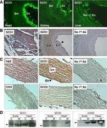
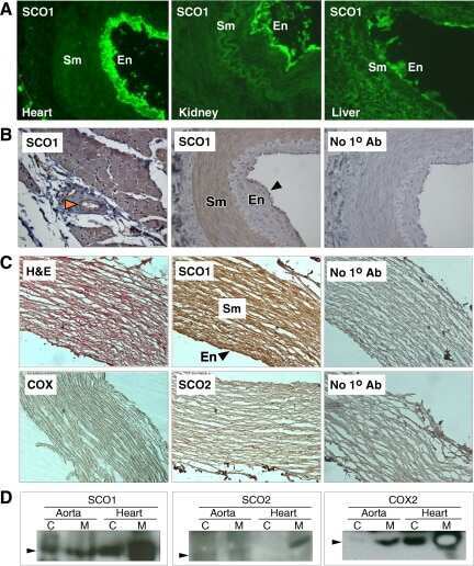
Unexpected vascular enrichment of SCO1 over SCO2 in mammalian tissues: implications for human mitochondrial disease.
Brosel S, Yang H, Tanji K, Bonilla E, Schon EA
The American journal of pathology 2010 Nov;177(5):2541-8
The American journal of pathology 2010 Nov;177(5):2541-8
No comments: Submit comment
Supportive validation
- Submitted by
- Invitrogen Antibodies (provider)
- Main image
- Experimental details
- NULL
- Submitted by
- Invitrogen Antibodies (provider)
- Main image

- Experimental details
- NULL
- Submitted by
- Invitrogen Antibodies (provider)
- Main image

- Experimental details
- NULL
- Submitted by
- Invitrogen Antibodies (provider)
- Main image
- Experimental details
- NULL

- Submitted by
- Invitrogen Antibodies (provider)
- Main image
- Experimental details
- NULL

- Submitted by
- Invitrogen Antibodies (provider)
- Main image
- Experimental details
- NULL
- Submitted by
- Invitrogen Antibodies (provider)
- Main image

- Experimental details
- NULL

- Submitted by
- Invitrogen Antibodies (provider)
- Main image
- Experimental details
- NULL
- Submitted by
- Invitrogen Antibodies (provider)
- Main image

- Experimental details
- NULL
- Submitted by
- Invitrogen Antibodies (provider)
- Main image

- Experimental details
- NULL
- Submitted by
- Invitrogen Antibodies (provider)
- Main image
- Experimental details
- NULL
- Submitted by
- Invitrogen Antibodies (provider)
- Main image
- Experimental details
- NULL

- Submitted by
- Invitrogen Antibodies (provider)
- Main image
- Experimental details
- NULL
- Submitted by
- Invitrogen Antibodies (provider)
- Main image
- Experimental details
- NULL
- Submitted by
- Invitrogen Antibodies (provider)
- Main image

- Experimental details
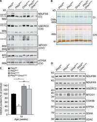
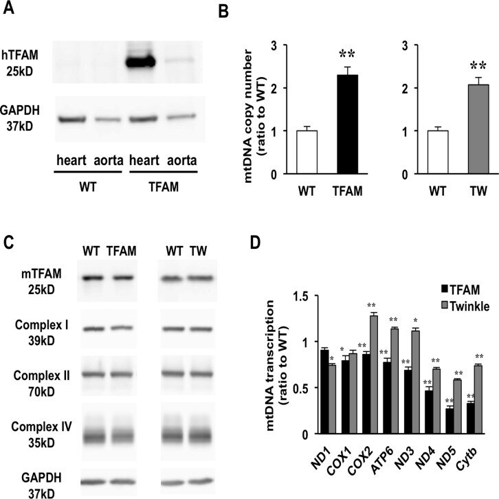
- Fig 1 Characterization of TFAM and Twinkle (TW) mice. (A) Expression of human TFAM (hTFAM) in left ventricle (LV) and aorta in TFAM and wild-type (WT) control mice. (B) mtDNA copy number in myocardium from TFAM and TW mice by real-time PCR (n = 4). (C) Expression of endogenous murine TFAM (mTFAM) and mitochondrial complex proteins in LV of TFAM and TW mice. (D) Transcription of mtDNA-encoded genes in TFAM and TW mice (n = 6). Data are expressed as mean +- SEM. * P < 0.05 vs. WT, ** P < 0.01 vs. WT, analyzed by Student's t- test.
- Submitted by
- Invitrogen Antibodies (provider)
- Main image
- Experimental details
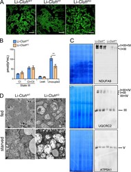
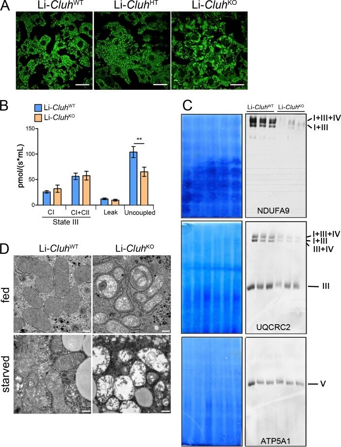
- Figure 5. Liver-specific Cluh deletion affects mitochondrial distribution and structure, assembled respiratory supercomplexes, and respiratory capacity. (A) Representative confocal images of livers of 8-wk-old mice of the indicated genotypes. To analyze mitochondrial morphology, mice were crossed with a stop-mito-YFP reporter line activated by Cre recombination. n = 4. Bars, 5 um. (B) Oxygen consumption of mitochondria isolated from livers of 8-wk-old mice. State III respiration was measured in the presence of pyruvate, malate, glutamate, and ADP (complex I [CI]), followed by addition of succinate (complex I + complex II [CI + CII]). The proton leak was measured after addition of oligomycin, whereas maximal respiration was assessed by CCCP titration. n = 5. Graph shows means +- SEM. **, P
- Submitted by
- Invitrogen Antibodies (provider)
- Main image

- Experimental details
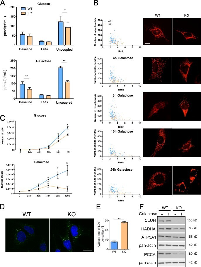
- Figure 7. Cluh -deficient MEFs mimic liver phenotypes. (A) Oxygen consumption of intact MEFs cultured in glucose or galactose medium. The proton leak was measured after the addition of oligomycin, whereas maximal respiration was assessed by CCCP titration. n >= 4. (B) Mitochondrial morphology in MEFs transfected with mito-mCherry. Graphs show the mean aspect ratio (area/perimeter) on the x axis and the number of mitochondria on the y axis for individual cells from three independent experiments. Right panels show representative images of mitochondrial morphology at the indicated time points. Bar, 12 um. (C) Growth curves of MEFs cultured in glucose or galactose medium during five consecutive days. n = 3. (D) Representative images of LD staining in MEFs grown in glucose medium. Nuclei were stained with DAPI (blue), and LDs were stained with BODIPY 493/503 (green). Bar, 20 um. (E) Quantification of LD staining shown in D. 50 cells were analyzed per genotype per experiment. Graph shows the mean area of LDs per cell. n = 3. (A, C, and E) Error bars are means +- SEM. *, P
- Submitted by
- Invitrogen Antibodies (provider)
- Main image
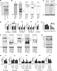
- Experimental details
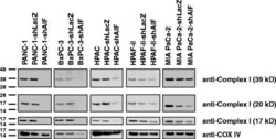
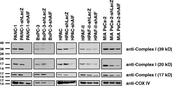
- Fig. 5 AIF selectively controls respiratory chain protein expression in pancreatic cancer cells. Following suppression of AIF, respiratory chain status was assessed by immunoblot analysis of complex I (39-, 20-, and 17-kDa subunits) and COX IV
- Submitted by
- Invitrogen Antibodies (provider)
- Main image

- Experimental details
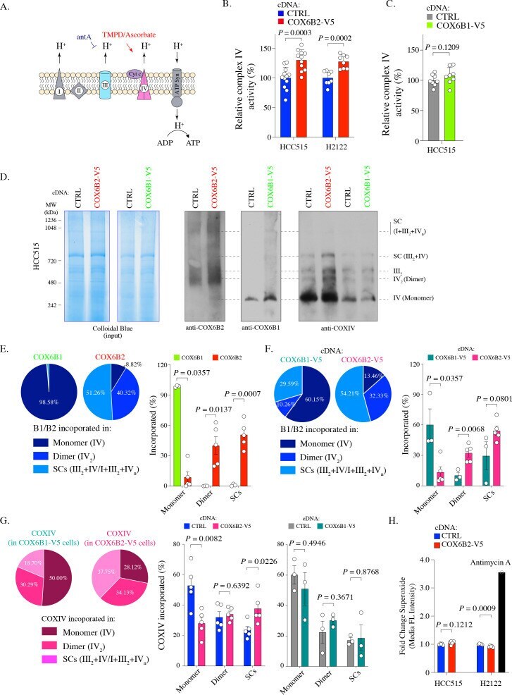
- Figure 3. COX6B2 enhances complex IV activity of OXPHOS without increasing ROS production. ( A ) Schematic of complex IV activity measurements in isolated mitochondria. Details are described in Material and methods. antA, Antimycin A; Cyt c, Cytochrome c; ATP Syn, ATP synthase. ( B ) Complex IV activity in indicated cell lines using TMPD/ascorbate as a substrate. Bars represent mean + SEM (n >= 8). p-Value calculated by Student's t -test. ( C ) As in ( B ) in HCC515 cell lines expressing CTRL and COX6B1-V5 cDNA. Bars represent mean + SEM (n = 8). p-Value calculated by Student's t -test. ( D ) Indicated lysates from HCC515 cells were run on a BN-PAGE gel and stained with Colloidal Blue (left two panels) or immunoblotted with anti-COX6B2, anti-COX6B1 and anti-COXIV (right panels). Representative image of n >= 3. MW markers are indicated to identify different complexes (). ( E ) Left: Distribution of COX6B1 and COX6B2 incorporated in complex IV monomers, dimers or supercomplexes as detected by BN-PAGE in ( D ). Right: Bars represent mean +- SEM (n >= 3) based on quantification of bands in ( D ). In monomeric complex IV, p value calculated by Mann-Whitney test whereas others calculated by Student's t -test. ( F ) Left: Distribution of COX6B1/COX6B2 incorporated in monomeric complex IV, dimeric complex IV and supercomplexes based on BN-PAGE in COX6B2-V5 or COX6B1-V5 overexpressing cell lines ( D ). Right: Bars represent mean +- SEM (n >= 3) based on quantification of bands in ( D
- Submitted by
- Invitrogen Antibodies (provider)
- Main image

- Experimental details
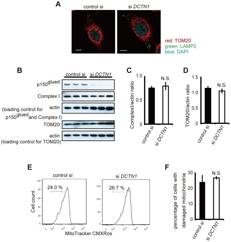
- Figure 4 Depletion of p150 glued does not induce damaged mitochondria accumulation. (A) Control siRNA or DCTN1 siRNA transfected cells were fixed and co-stained with antibodies against LAMP2 (green) and TOM20 (red), and analyzed using confocal microscopy. Bars, 10 mum. (B) Control siRNA or DCTN1 siRNA transfected HeLa cells were analyzed by immunoblotting with antibodies against complex I, TOM20, and actin. (C, D) Densitometry analysis of complex I (C) and TOM20 (D) levels relative to actin was performed. (E, F) DCTN1 siRNA transfected HeLa cells were incubated with Mitotracker-Red CMXRos and intracellular fluorescence intensity was measured by flow cytometry. The histograms of MitoTracker-Red CMXRos fluorescence (E) and the percentages of cells with reduced mitochondrial potentials (F) are shown. The error bar indicates each standard deviation. Statistics are from three independent experiments: N.S., not significant.
- Submitted by
- Invitrogen Antibodies (provider)
- Main image
- Experimental details
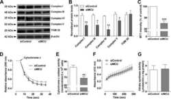
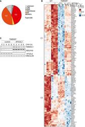
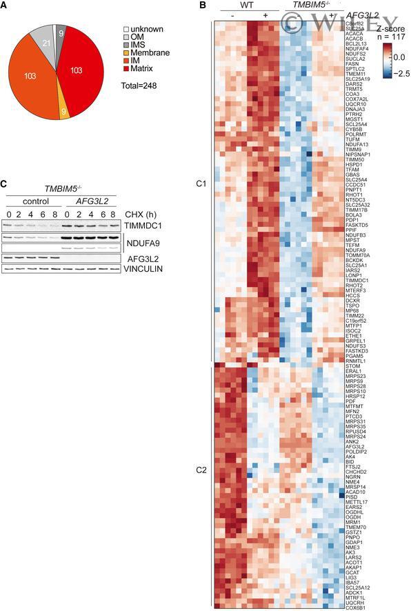
- 5 Figure Loss of TMBIM5 promotes proteolysis by AFG3L2 A Pie chart showing the localization of proteins accumulating at decreased levels in TMBIM5 -/- HeLa cells. Localization according to MitoCarta 3.0. OM, outer membrane; IMS, intermembrane space; IM, inner membrane. B Alterations in mitochondrial proteome in WT and TMBIM5 -/- HeLa cells upon depletion of AFG3L2. WT and TMBIM5 -/- HeLa cells were transfected with scrambled siRNA (control) or siRNA targeting AFG3L2 for 48 h. Proteins, which were downregulated in the absence of TMBIM5 and whose steady-state levels were altered upon depletion of AFG3L2, were identified using a two-way ANOVA. An interaction P -value of 0.01 was used as a cutoff. Log2-transformed LFQ intensities of MitoCarta 3.0 proteins were Z -Score normalized and visualized in hierarchical cluster analysis (Euclidean distance, complete method). The dendrogram is omitted. Proteins accumulating upon depletion of AFG3L2 (cluster 1, C1) and proteins whose steady-state levels are decreased upon depletion of AFG3L2 (cluster 2, C2) are shown. See also Dataset EV6. C Representative immunoblot of TMBIM5 -/- HeLa cells transfected with scrambled siRNA (control) or siRNA targeting AFG3L2 for 48 h. Samples were treated with the protein synthesis inhibitor cycloheximide (CHX; 10 mug/ml) and collected at the indicated time points ( n = 3 independent experiments). Data information: See also Dataset EV6. Source data are available online for this figure.
- Submitted by
- Invitrogen Antibodies (provider)
- Main image

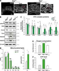
- Experimental details
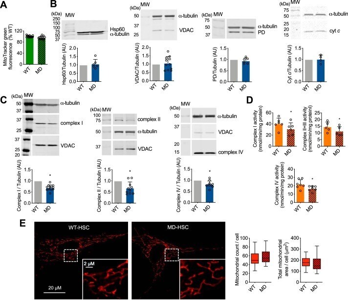
- Figure 4. Loss of merlin expression in human Schwann cells leads to decreased mitochondrial oxidative phosphorylation complex levels and activity. A, the mitochondrial content of human WT- and MD-Schwann cells was determined after 30 min incubation with the mitochondrial membrane potential-independent probe MitoTracker Green (200 n m ) and expressed as percentage of WT-Schwann cells ( n = 3 with 4 replicates). B and C, representative IR Western blots of mitochondrial proteins in human WT- and MD-Schwann cells: B, Hsp60, VDAC, PD, and cytochrome c (cyt c ); and C, complex I (NDUFA9 subunit), complex II (SDHA subunit), and complex IV (subunit COX IV). Below , quantitation of the corresponding bands normalized against alpha-tubulin and expressed relative to WT-Schwann cells ( n = 4-12). D, mitochondrial complex I, II + III, and IV activities were assessed in disrupted isolated mitochondria of WT- and MD-Schwann cells. Complex I activity was measured at 340 nm by the rotenone-sensitive reduction of ubiquinone-1 in the presence of potassium cyanide and NADH. Complex II + III activity was determined by the antimycin A-sensitive reduction of cytochrome c at 550 nm in the presence of potassium cyanide and succinate. Complex IV activity was determined by monitoring the potassium cyanide-sensitive oxidation of cytochrome c at 550 nm. Activities are expressed as nanomole/min/mg of protein; columns represent the mean +- S.D. ( n = 6-7). E, representative images