Explore
 Validate
 Learn
Learn Western blot
Antibody data
- Antibody Data
- Antigen structure
- References [0]
- Comments [0]
- Validations
- Western blot [3]
- Immunocytochemistry [1]
Submit
Validation data
Reference
Comment
Report error
- Product number
- PA5-67275 - Provider product page

- Provider
- Invitrogen Antibodies
- Product name
- NDUFA9 Polyclonal Antibody
- Antibody type
- Polyclonal
- Antigen
- Recombinant full-length protein
- Description
- Immunogen sequence: AQLSKEAGVEK FIHVSHLNAN IKSSSRYLRN KAVGEKVVRD AFPEAIIVKP SDIFGREDRF LNSFASMHRF GPIPL Highest antigen sequence identity to the following orthologs - mouse 74%, rat 70%.
- Reactivity
- Human
- Host
- Rabbit
- Isotype
- IgG
- Vial size
- 100 µL
- Concentration
- 0.05 mg/mL
- Storage
- Store at 4°C short term. For long term storage, store at -20°C, avoiding freeze/thaw cycles.
No comments: Submit comment
Supportive validation
- Submitted by
- Invitrogen Antibodies (provider)
- Main image
- Experimental details
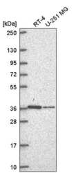
- Western blot analysis of NDUFA9 in human cell line RT-4 and human cell line U-251 MG. Samples were probed using a NDUFA9 Polyclonal Antibody (Product # PA5-67275).

- Submitted by
- Invitrogen Antibodies (provider)
- Main image
- Experimental details
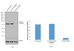
- Knockdown of NDUFA9 was achieved by transfecting Hep G2 cells with NDUFA9 specific siRNAs (Silencer® select Product # s39346, s9347). Western blot analysis (Fig. a) was performed using membrane enriched cell extracts from the NDUFA9 knockdown cells (lane 3), non-specific scrambled siRNA transfected cells (lane 2) and untransfected cells (lane 1). The blot was probed NDUFA9 Polyclonal Antibody (Product # PA5-67275, 1:500 dilution) and Goat anti-Rabbit IgG (H+L) Superclonal™ Secondary Antibody, HRP conjugate (Product # A27036, 0.25 ug/ml, 1:4000 dilution). Densitometric analysis of this western blot is shown in histogram (Fig. b). Decrease in signal upon siRNA mediated knock down confirms that antibody is specific to NDUFA9.

- Submitted by
- Invitrogen Antibodies (provider)
- Main image
- Experimental details
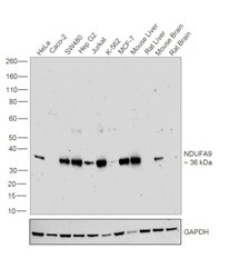
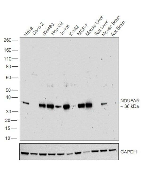
- Western blot was performed using NDUFA9 Polyclonal Antibody (Product # PA5-67275) and a 36 kDa band corresponding to NDUFA9 was observed across cell lines and tissues tested. Membrane enriched cell extracts (30 µg lysate) of HeLa (Lane 1), Caco-2 (Lane 2), SW480 (Lane 3), Hep G2 (Lane 4), Jurkat (Lane 5), K-562 (Lane 6), MCF-7 (Lane 7), Mouse Liver (Lane 8), Rat Liver (Lane 9), Mouse Brain (Lane 10) and Rat Brain (Lane 11) were electrophoresed using Novex® NuPAGE® 12 % Bis-Tris gel (Product # NP0342BOX). Resolved proteins were then transferred onto a nitrocellulose membrane (Product # IB23001) by iBlot® 2 Dry Blotting System (Product # IB21001). The blots were probed with the primary antibody (1:500 dilution) and detected by chemiluminescence with Goat Anti-Rabbit IgG Secondary Antibody, HRP conjugate (Product # A27036, 1:4000 dilution) using the iBright FL 1000 (Product # A32752). Chemiluminescent detection was performed using Novex® ECL Chemiluminescent Substrate Reagent Kit (Product # WP20005).
Supportive validation
- Submitted by
- Invitrogen Antibodies (provider)
- Main image

- Experimental details
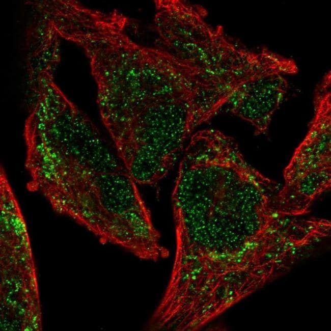
- Immunofluorescent staining of NDUFA9 in human cell line RH-30 shows localization to nucleoplasm and mitochondria. Samples were probed using a NDUFA9 Polyclonal Antibody (Product # PA5-67275).