 Explore
 Validate
 Learn
Learn Western blot
 Immunocytochemistry
 Immunohistochemistry
Antibody data
- Antibody Data
- Antigen structure
- References [0]
- Comments [0]
- Validations
- Western blot [1]
- Immunocytochemistry [2]
Submit
Validation data
Reference
Comment
Report error
- Product number
- LS-C773940 - Provider product page

- Provider
- LSBio
- Product name
- GABRA5 Antibody (clone S415-24, Biotin) LS-C773940
- Antibody type
- Monoclonal
- Description
- Protein G purified
- Reactivity
- Human, Mouse, Rat
- Host
- Mouse
- Conjugate
- Biotin
- Isotype
- IgG
- Antibody clone number
- S415-24
- Storage
- Store at -20°C.
No comments: Submit comment
Enhanced validation
- Submitted by
- LSBio (provider)
- Enhanced method
- Genetic validation
- Main image

- Experimental details
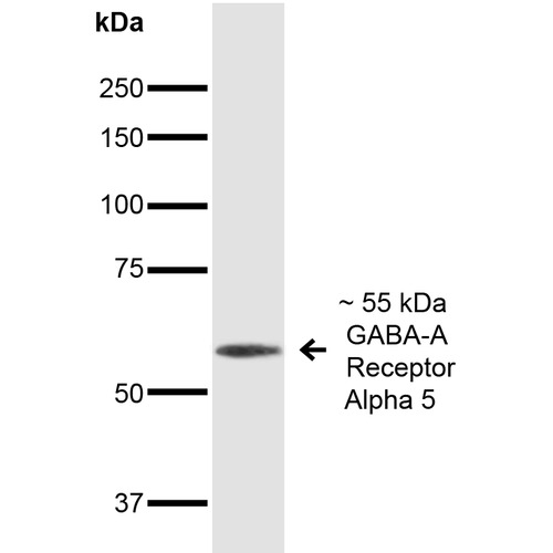
- Western Blot analysis of Mouse Brain showing detection of ~55 kDa GABA A Receptor Alpha 5 protein using Mouse Anti-GABA A Receptor Alpha 5 Monoclonal Antibody, Clone S415-24. Lane 1: MW Ladder. Lane 2: Mouse Brain. Load: 20 µg. Primary Antibody: Mouse Anti-GABA A Receptor Alpha 5 Monoclonal Antibody at 1:1000 for 16 hours at 4°C. Secondary Antibody: Goat Anti-Mouse IgG: HRP at 1:200 for 1 hour at RT. Predicted/Observed Size: ~55 kDa.
Supportive validation
- Submitted by
- LSBio (provider)
- Enhanced method
- Genetic validation
- Main image
- Experimental details
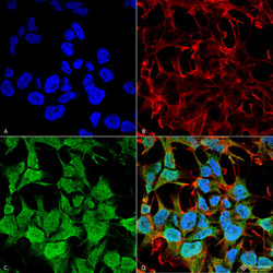
- Immunocytochemistry/Immunofluorescence analysis using Mouse Anti-GABA-A Receptor Alpha5 Monoclonal Antibody, Clone S415-24. Tissue: Neuroblastoma cell line (SK-N-BE). Species: Human. Fixation: 4% Formaldehyde for 15 min at RT. Primary Antibody: Mouse Anti-GABA-A Receptor Alpha5 Monoclonal Antibody at 1:100 for 60 min at RT. Secondary Antibody: Goat Anti-Mouse ATTO 488 at 1:100 for 60 min at RT. Counterstain: Phalloidin Texas Red F-Actin stain; DAPI (blue) nuclear stain at 1:1000, 1:5000 for 60min RT, 5min RT. Localization: Cell Junction, Synapse, Postsynaptic Cell Membrane, Cell Membrane. Magnification: 60X. (A) DAPI (blue) nuclear stain. (B) Phalloidin Texas Red F-Actin stain. (C) GABA-A Receptor Alpha5 Antibody. (D) Composite.

- Submitted by
- LSBio (provider)
- Main image
- Experimental details
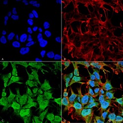
- Immunocytochemistry/Immunofluorescence analysis using Mouse Anti-GABA-A Receptor Alpha5 Monoclonal Antibody, Clone S415-24. Tissue: Neuroblastoma cell line (SK-N-BE). Species: Human. Fixation: 4% Formaldehyde for 15 min at RT. Primary Antibody: Mouse Anti-GABA-A Receptor Alpha5 Monoclonal Antibody at 1:100 for 60 min at RT. Secondary Antibody: Goat Anti-Mouse ATTO 488 at 1:100 for 60 min at RT. Counterstain: Phalloidin Texas Red F-Actin stain; DAPI (blue) nuclear stain at 1:1000, 1:5000 for 60min RT, 5min RT. Localization: Cell Junction, Synapse, Postsynaptic Cell Membrane, Cell Membrane. Magnification: 60X. (A) DAPI (blue) nuclear stain. (B) Phalloidin Texas Red F-Actin stain. (C) GABA-A Receptor Alpha5 Antibody. (D) Composite.
