Explore
 Validate
 Learn
Learn Western blot
Antibody data
- Antibody Data
- Antigen structure
- References [0]
- Comments [0]
- Validations
- Western blot [1]
- Immunocytochemistry [1]
- Immunohistochemistry [3]
Submit
Validation data
Reference
Comment
Report error
- Product number
- TA328692 - Provider product page

- Provider
- OriGene
- Product name
- Rabbit Polyclonal Anti-HCN2
- Antibody type
- Polyclonal
- Description
- Rabbit Polyclonal Anti-HCN2
- Host
- Rabbit
- Conjugate
- Unconjugated
- Epitope
- HCN2
- Antibody clone number
- NULL
- Vial size
- 200 µl
- Concentration
- NULL
No comments: Submit comment
Supportive validation
- Submitted by
- OriGene (provider)
- Main image

- Experimental details
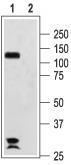
- Western blot analysis of rat brain membranes: 1. Anti-HCN2 antibody, (1:200). 2. Anti-HCN2 antibody, preincubated with the control peptide antigen.
- Validation comment
- WB
Supportive validation
- Submitted by
- OriGene (provider)
- Main image
- Experimental details
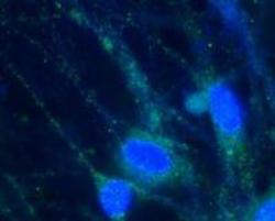
- Expression of HCN2 in rat DRG primary culture. Immunocytochemical staining of paraformaldehyde-fixed and permeabilized rat dorsal root ganglion (DRG) primary culture using Anti-HCN2 antibody, (1:100), (green). Cells were stained with Anti-HCN2 antibody followed by goat anti-rabbit-AlexaFluor-488 secondary antibody. Nuclear staining of cells using the cell-permeable DNA dye Hoechst 33342 (blue).
- Validation comment
- IF

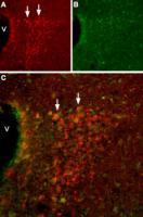
Supportive validation
- Submitted by
- OriGene (provider)
- Main image
- Experimental details
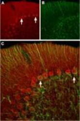
- Expression of HCN2 in rat cerebellum. Immunochistochemical staining of rat cerebellum frozen sections using Anti-HCN2 antibody. A. HCN2 (red) appears in Purkinje cells (arrows). B. Staining of astrocytes with mouse anti-glial fibrillary acidic protein (GFAP, green demonstrates the restriction of HCN2 to neuronal cell bodies. C. Confocal merge of HCN2 and GFAP images demonstrates the respective localization of these proteins.
- Validation comment
- IHC

- Submitted by
- OriGene (provider)
- Main image
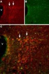
- Experimental details
- Expression of HCN2 in mouse hypothalamus. Immunohistochemical staining of mouse hypothalamus using Anti-HCN2 antibody. A. HCN2 (red) appears in cells of the paraventricular nucleus (PVN, arrows). B. Staining of paraventricular nerve cells with mouse anti-calcium binding protein (CBD28k, green). C. Confocal merge of HCN2 and CBD28k demonstrates some co-localization. V = Third ventricle.
- Validation comment
- IHC
- Submitted by
- OriGene (provider)
- Main image
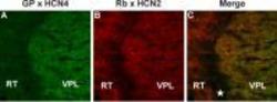
- Experimental details
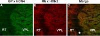
- IHC staining of mouse thalamus frozen section using guinea pig Anti-HCN4 antibody and rabbit Anti-HCN2 antibody. A. Staining of HCN4 (green) appears in the ventral posterior thalamic nucleus (VPL). B. In the same section as in A, staining of HCN2 (red) appears in the ventral posterior thalamic nucleus (VPL) and also in the reticular thalamic nucleus (RT). The area between these thalamic nuclei (star) is white matter and neither protein is expressed in that region. C. Merged images of A and B.
- Validation comment
- IHC