Explore
 Validate
 Learn
Learn Western blot
 Immunohistochemistry
Antibody data
- Antibody Data
- Antigen structure
- References [3]
- Comments [0]
- Validations
- Immunohistochemistry [1]
Submit
Validation data
Reference
Comment
Report error
- Product number
- A02804 - Provider product page

- Provider
- Boster Biological Technology
- Product name
- Anti-HCN2 Antibody Picoband™
- Antibody type
- Polyclonal
- Description
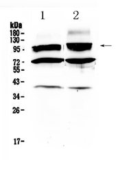
- Polyclonal antibody for HCN2 detection. Host: Rabbit.Size: 100μg/vial. Tested applications: IHC-P. Reactive species: Human. HCN2 information: Molecular Weight: 96950 MW; Subcellular Localization: Cell membrane ; Multi-pass membrane protein ; Tissue Specificity: Highly expressed throughout the brain. Detected at low levels in heart.
- Reactivity
- Human, Mouse, Rat
- Host
- Rabbit
- Vial size
- 100μg/vial
- Concentration
- Add 0.2ml of distilled water will yield a concentration of 500ug/ml.
- Storage
- At -20°C for one year. After reconstitution, at 4°C for one month. It can also be aliquoted and stored frozen at -20°C for a longer time. Avoid repeated freezing and thawing.
- Handling
- Add 0.2ml of distilled water will yield a concentration of 500ug/ml.
Submitted references The HCN domain is required for HCN channel cell-surface expression and couples voltage- and cAMP-dependent gating mechanisms.
Spironolactone diminishes spontaneous ventricular premature beats by reducing HCN4 protein expression in rats with myocardial infarction.
Dynamic changes in HCN2, HCN4, KCNE1, and KCNE2 expression in ventricular cells from acute myocardial infarction rat hearts.
Wang ZJ, Blanco I, Hayoz S, Brelidze TI
The Journal of biological chemistry 2020 Jun 12;295(24):8164-8173
The Journal of biological chemistry 2020 Jun 12;295(24):8164-8173
Spironolactone diminishes spontaneous ventricular premature beats by reducing HCN4 protein expression in rats with myocardial infarction.
Song T, Yang J, Yao Y, Li H, Chen Y, Zhang J, Huang C
Molecular medicine reports 2011 May-Jun;4(3):569-73
Molecular medicine reports 2011 May-Jun;4(3):569-73
Dynamic changes in HCN2, HCN4, KCNE1, and KCNE2 expression in ventricular cells from acute myocardial infarction rat hearts.
Xia S, Wang Y, Zhang Y, Deng SB, Du JL, Wang XC, She Q
Biochemical and biophysical research communications 2010 May 7;395(3):330-5
Biochemical and biophysical research communications 2010 May 7;395(3):330-5
No comments: Submit comment
Supportive validation
- Submitted by
- Boster Biological Technology (provider)
- Main image
- Experimental details
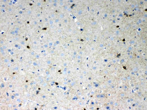
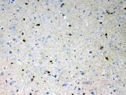
- IHC analysis of HCN2 using anti- HCN2 antibody (A02804). HCN2 was detected in paraffin-embedded section of rat brain tissues. Heat mediated antigen retrieval was performed in citrate buffer (pH6, epitope retrieval solution) for 20 mins. The tissue section was blocked with 10% goat serum. The tissue section was then incubated with 1μg/ml rabbit anti- HCN2 Antibody (A02804) overnight at 4°C. Biotinylated goat anti-rabbit IgG was used as secondary antibody and incubated for 30 minutes at 37°C. The tissue section was developed using Strepavidin-Biotin-Complex (SABC)(Catalog # SA1022) with DAB as the chromogen.
- Additional image