Explore
 Validate
 Learn
Learn Western blot
 Immunocytochemistry
Antibody data
- Antibody Data
- Antigen structure
- References [11]
- Comments [0]
- Validations
- Immunocytochemistry [4]
- Flow cytometry [3]
- Other assay [1]
Submit
Validation data
Reference
Comment
Report error
- Product number
- MA5-13493 - Provider product page

- Provider
- Invitrogen Antibodies
- Product name
- CD155 Monoclonal Antibody (D171)
- Antibody type
- Monoclonal
- Antigen
- Other
- Description
- MA5-13493 targets CD155 in Western blot, FACS and ICC/IF applications and shows reactivity with Human, Mouse and Non-human primate samples. The MA5-13493 immunogen is HeLa cells.
- Reactivity
- Human, Mouse
- Host
- Mouse
- Isotype
- IgG
- Antibody clone number
- D171
- Vial size
- 500 μL
- Concentration
- 0.2 mg/mL
- Storage
- 4°C
Submitted references Inhibition of clathrin-mediated endocytosis by knockdown of AP-2 leads to alterations in the plasma membrane proteome.
High expression of soluble CD155 in estrogen receptor-negative breast cancer.
Innate immune activating ligand SUMOylation affects tumor cell recognition by NK cells.
Inhibition of Necl-5 (CD155/PVR) reduces glioblastoma dispersal and decreases MMP-2 expression and activity.
NK cells recognize and lyse Ewing sarcoma cells through NKG2D and DNAM-1 receptor dependent pathways.
CD155/PVR enhances glioma cell dispersal by regulating adhesion signaling and focal adhesion dynamics.
DNAM-1 and PVR regulate monocyte migration through endothelial junctions.
Receptor (CD155)-dependent endocytosis of poliovirus and retrograde axonal transport of the endosome.
Identification of secreted CD155 isoforms.
Identification of secreted CD155 isoforms.
Overexpression of the CD155 gene in human colorectal carcinoma.
Tobys D, Kowalski LM, Cziudaj E, Müller S, Zentis P, Pach E, Zigrino P, Blaeske T, Höning S
Traffic (Copenhagen, Denmark) 2021 Jan;22(1-2):6-22
Traffic (Copenhagen, Denmark) 2021 Jan;22(1-2):6-22
High expression of soluble CD155 in estrogen receptor-negative breast cancer.
Iguchi-Manaka A, Okumura G, Ichioka E, Kiyomatsu H, Ikeda T, Bando H, Shibuya A, Shibuya K
Breast cancer (Tokyo, Japan) 2020 Jan;27(1):92-99
Breast cancer (Tokyo, Japan) 2020 Jan;27(1):92-99
Innate immune activating ligand SUMOylation affects tumor cell recognition by NK cells.
Zitti B, Molfetta R, Fionda C, Quatrini L, Stabile H, Lecce M, de Turris V, Ricciardi MR, Petrucci MT, Cippitelli M, Gismondi A, Santoni A, Paolini R
Scientific reports 2017 Sep 5;7(1):10445
Scientific reports 2017 Sep 5;7(1):10445
Inhibition of Necl-5 (CD155/PVR) reduces glioblastoma dispersal and decreases MMP-2 expression and activity.
Enloe BM, Jay DG
Journal of neuro-oncology 2011 Apr;102(2):225-35
Journal of neuro-oncology 2011 Apr;102(2):225-35
NK cells recognize and lyse Ewing sarcoma cells through NKG2D and DNAM-1 receptor dependent pathways.
Verhoeven DH, de Hooge AS, Mooiman EC, Santos SJ, ten Dam MM, Gelderblom H, Melief CJ, Hogendoorn PC, Egeler RM, van Tol MJ, Schilham MW, Lankester AC
Molecular immunology 2008 Sep;45(15):3917-25
Molecular immunology 2008 Sep;45(15):3917-25
CD155/PVR enhances glioma cell dispersal by regulating adhesion signaling and focal adhesion dynamics.
Sloan KE, Stewart JK, Treloar AF, Matthews RT, Jay DG
Cancer research 2005 Dec 1;65(23):10930-7
Cancer research 2005 Dec 1;65(23):10930-7
DNAM-1 and PVR regulate monocyte migration through endothelial junctions.
Reymond N, Imbert AM, Devilard E, Fabre S, Chabannon C, Xerri L, Farnarier C, Cantoni C, Bottino C, Moretta A, Dubreuil P, Lopez M
The Journal of experimental medicine 2004 May 17;199(10):1331-41
The Journal of experimental medicine 2004 May 17;199(10):1331-41
Receptor (CD155)-dependent endocytosis of poliovirus and retrograde axonal transport of the endosome.
Ohka S, Matsuda N, Tohyama K, Oda T, Morikawa M, Kuge S, Nomoto A
Journal of virology 2004 Jul;78(13):7186-98
Journal of virology 2004 Jul;78(13):7186-98
Identification of secreted CD155 isoforms.
Baury B, Masson D, McDermott BM Jr, Jarry A, Blottière HM, Blanchardie P, Laboisse CL, Lustenberger P, Racaniello VR, Denis MG
Biochemical and biophysical research communications 2003 Sep 12;309(1):175-82
Biochemical and biophysical research communications 2003 Sep 12;309(1):175-82
Overexpression of the CD155 gene in human colorectal carcinoma.
Masson D, Jarry A, Baury B, Blanchardie P, Laboisse C, Lustenberger P, Denis MG
Gut 2001 Aug;49(2):236-40
Gut 2001 Aug;49(2):236-40
No comments: Submit comment
Supportive validation
- Submitted by
- Invitrogen Antibodies (provider)
- Main image
- Experimental details
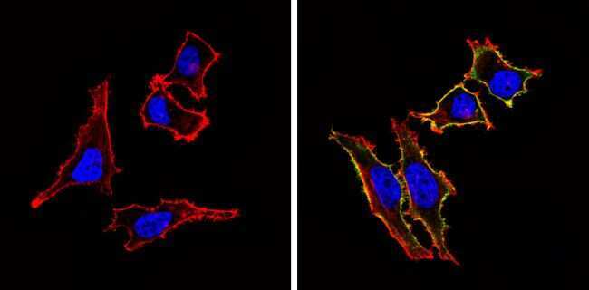
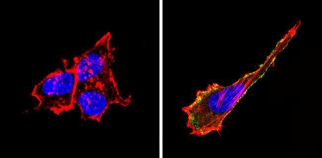
- Immunofluorescent analysis of CD155 (green) showing staining in the membrane of Hela cells (right) compared to a negative control without primary antibody (left). Formalin-fixed cells were permeabilized with 0.1% Triton X-100 in TBS for 5-10 minutes and blocked with 3% BSA-PBS for 30 minutes at room temperature. Cells were probed with a CD155 monoclonal antibody (Product # MA5-13493) in 3% BSA-PBS at a dilution of 1:20 and incubated overnight at 4 ºC in a humidified chamber. Cells were washed with PBST and incubated with a DyLight-conjugated secondary antibody in PBS at room temperature in the dark. F-actin (red) was stained with a fluorescent red phalloidin and nuclei (blue) were stained with Hoechst or DAPI. Images were taken at a magnification of 60x.
- Submitted by
- Invitrogen Antibodies (provider)
- Main image
- Experimental details
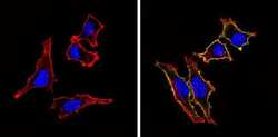
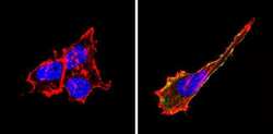
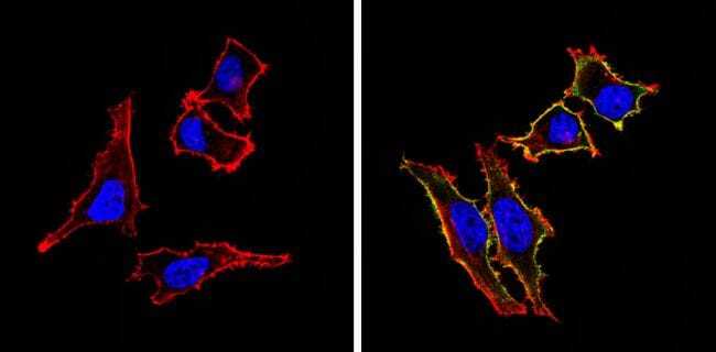
- Immunofluorescent analysis of CD155 (green) showing staining in the membrane of U-87 MG cells (right) compared to a negative control without primary antibody (left). Formalin-fixed cells were permeabilized with 0.1% Triton X-100 in TBS for 5-10 minutes and blocked with 3% BSA-PBS for 30 minutes at room temperature. Cells were probed with a CD155 monoclonal antibody (Product # MA5-13493) in 3% BSA-PBS at a dilution of 1:20 and incubated overnight at 4 ºC in a humidified chamber. Cells were washed with PBST and incubated with a DyLight-conjugated secondary antibody in PBS at room temperature in the dark. F-actin (red) was stained with a fluorescent red phalloidin and nuclei (blue) were stained with Hoechst or DAPI. Images were taken at a magnification of 60x.
- Submitted by
- Invitrogen Antibodies (provider)
- Main image
- Experimental details
- Immunofluorescent analysis of CD155 (green) showing staining in the membrane of U-87 MG cells (right) compared to a negative control without primary antibody (left). Formalin-fixed cells were permeabilized with 0.1% Triton X-100 in TBS for 5-10 minutes and blocked with 3% BSA-PBS for 30 minutes at room temperature. Cells were probed with a CD155 monoclonal antibody (Product # MA5-13493) in 3% BSA-PBS at a dilution of 1:20 and incubated overnight at 4 ºC in a humidified chamber. Cells were washed with PBST and incubated with a DyLight-conjugated secondary antibody in PBS at room temperature in the dark. F-actin (red) was stained with a fluorescent red phalloidin and nuclei (blue) were stained with Hoechst or DAPI. Images were taken at a magnification of 60x.

- Submitted by
- Invitrogen Antibodies (provider)
- Main image

- Experimental details
- Immunofluorescent analysis of CD155 (green) showing staining in the membrane of Hela cells (right) compared to a negative control without primary antibody (left). Formalin-fixed cells were permeabilized with 0.1% Triton X-100 in TBS for 5-10 minutes and blocked with 3% BSA-PBS for 30 minutes at room temperature. Cells were probed with a CD155 monoclonal antibody (Product # MA5-13493) in 3% BSA-PBS at a dilution of 1:20 and incubated overnight at 4 ºC in a humidified chamber. Cells were washed with PBST and incubated with a DyLight-conjugated secondary antibody in PBS at room temperature in the dark. F-actin (red) was stained with a fluorescent red phalloidin and nuclei (blue) were stained with Hoechst or DAPI. Images were taken at a magnification of 60x.
Supportive validation
- Submitted by
- Invitrogen Antibodies (provider)
- Main image
- Experimental details
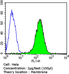
- Flow cytometry analysis of CD155 in Hela cells compared to an isotype control (blue). Cells were harvested, adjusted to a concentration of 1-5x10^6 cells/mL, fixed with 2% paraformaldehyde and washed with PBS. Cells were blocked with a 2% solution of BSA-PBS for 30 min at room temperature and incubated with a CD155 monoclonal antibody (Product # MA5-13493) at a dilution of 2 µg/test for 60 min at room temperature. Cells were then incubated for 40 min at room temperature in the dark using a Dylight 488-conjugated goat anti-mouse IgG (H+L) secondary antibody and re-suspended in PBS for FACS analysis.

- Submitted by
- Invitrogen Antibodies (provider)
- Main image

- Experimental details
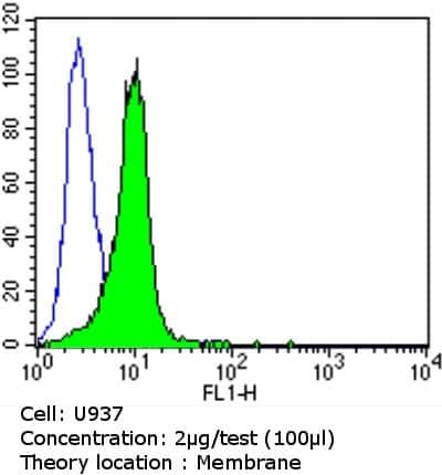
- Flow cytometry analysis of CD155 in U937 cells compared to an isotype control (blue). Cells were harvested, adjusted to a concentration of 1-5x10^6 cells/mL, fixed with 2% paraformaldehyde and washed with PBS. Cells were blocked with a 2% solution of BSA-PBS for 30 min at room temperature and incubated with a CD155 monoclonal antibody (Product # MA5-13493) at a dilution of 2 µg/test for 60 min at room temperature. Cells were then incubated for 40 min at room temperature in the dark using a Dylight 488-conjugated goat anti-mouse IgG (H+L) secondary antibody and re-suspended in PBS for FACS analysis.
- Submitted by
- Invitrogen Antibodies (provider)
- Main image

- Experimental details
- Flow cytometry analysis of CD155 in U937 cells compared to an isotype control (blue). Cells were harvested, adjusted to a concentration of 1-5x10^6 cells/mL, fixed with 2% paraformaldehyde and washed with PBS. Cells were blocked with a 2% solution of BSA-PBS for 30 min at room temperature and incubated with a CD155 monoclonal antibody (Product # MA5-13493) at a dilution of 2 µg/test for 60 min at room temperature. Cells were then incubated for 40 min at room temperature in the dark using a Dylight 488-conjugated goat anti-mouse IgG (H+L) secondary antibody and re-suspended in PBS for FACS analysis.

Supportive validation
- Submitted by
- Invitrogen Antibodies (provider)
- Main image
- Experimental details
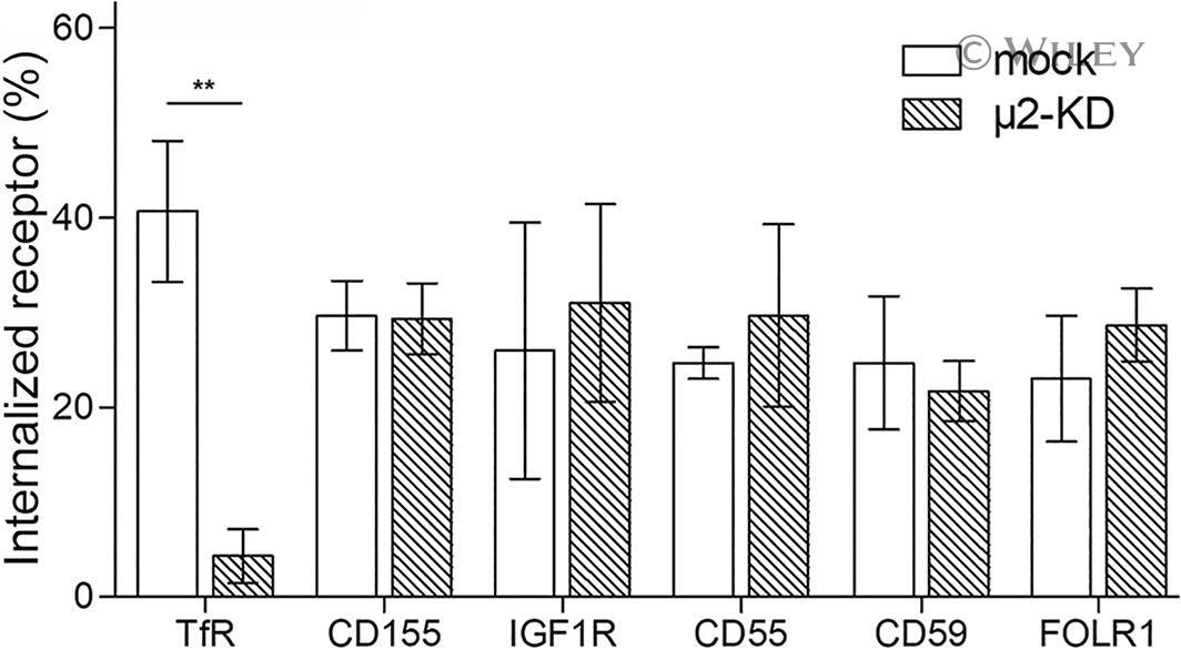
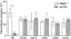
- 7 FIGURE Markers of clathrin-independent endocytosis are internalized by mu2 KD cells. Endocytosis of the indicated transmembrane (TfR; CD155; IGF1R) and GPI-anchored (CD55, CD59, FOLR1) proteins was determined by flow cytometry in mu2 KD and mock-treated cells. The fraction of internalized receptors represents the calculated ratio of fluorescence signal intensities obtained after labeling on ice with endocytosis for 2 minutes at 37degC and total labeling at the cell surface on ice without endocytosis. Note that CD155, IGF1R, CD55, CD59 and FOLR1 are published markers for clathrin-independent endocytosis. Error bars represent the SEM of data from three biological replicates (** P < .01; two-way ANOVA)