Explore
 Validate
 Learn
Learn Western blot
Antibody data
- Antibody Data
- Antigen structure
- References [0]
- Comments [0]
- Validations
- Western blot [3]
- Immunocytochemistry [1]
Submit
Validation data
Reference
Comment
Report error
- Product number
- AF2530 - Provider product page

- Provider
- R&D Systems
- Product name
- Human CD155/PVR Antibody
- Antibody type
- Polyclonal
- Description
- Antigen Affinity-purified. Detects human CD155/PVR in direct ELISAs and Western blots.
- Reactivity
- Human
- Host
- Goat
- Conjugate
- Unconjugated
- Antigen sequence
AAH15542- Isotype
- IgG
- Vial size
- 100 ug
- Concentration
- LYOPH
- Storage
- Use a manual defrost freezer and avoid repeated freeze-thaw cycles. 12 months from date of receipt, -20 to -70 °C as supplied. 1 month, 2 to 8 °C under sterile conditions after reconstitution. 6 months, -20 to -70 °C under sterile conditions after reconstitution.
No comments: Submit comment
Supportive validation
- Submitted by
- R&D Systems (provider)
- Main image

- Experimental details
- Detection of Human CD155/PVR by Western Blot. Western blot shows lysates of human heart tissue, HT1080 human fibrosarcoma cell line, and HUVEC human umbilical vein endothelial cells. PVDF membrane was probed with 0.25 µg/mL of Goat Anti-Human CD155/PVR Antigen Affinity-purified Polyclonal Antibody (Catalog # AF2530) followed by HRP-conjugated Anti-Goat IgG Secondary Antibody (Catalog # HAF017). A specific band was detected for CD155/PVR at approximately 75 kDa (as indicated). This experiment was conducted under reducing conditions and using Immunoblot Buffer Group 1.

- Submitted by
- R&D Systems (provider)
- Main image

- Experimental details
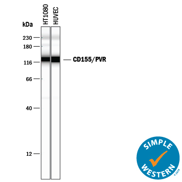
- Detection of Human CD155/PVR by Simple WesternTM. Simple Western lane view shows lysates of HT1080 human fibrosarcoma cell line and HUVEC human umbilical vein endothelial cells, loaded at 0.2 mg/mL. A specific band was detected for CD155/PVR at approximately 128 kDa (as indicated) using 2.5 µg/mL of Goat Anti-Human CD155/PVR Antigen Affinity-purified Polyclonal Antibody (Catalog # AF2530) followed by 1:50 dilution of HRP-conjugated Anti-Goat IgG Secondary Antibody (Catalog # HAF109). This experiment was conducted under reducing conditions and using the 12-230 kDa separation system.
- Submitted by
- R&D Systems (provider)
- Main image
- Experimental details
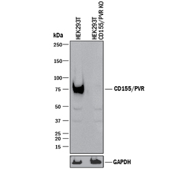
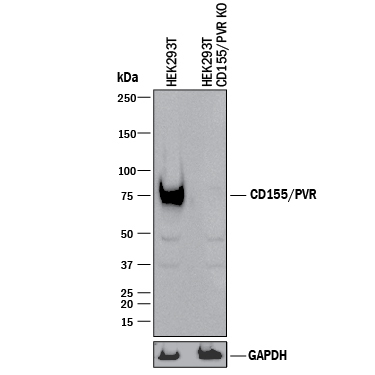
- Western Blot Shows Human CD155/PVR Specificity by Using Knockout Cell Line. Western blot shows lysates of HEK293T human embryonic kidney parental cell line and CD155/PVR knockout HEK293T cell line (KO). PVDF membrane was probed with 0.25 µg/mL of Goat Anti-Human CD155/PVR Antigen Affinity-purified Polyclonal Antibody (Catalog # AF2530) followed by HRP-conjugated Anti-Goat IgG Secondary Antibody (Catalog # HAF017). A specific band was detected for CD155/PVR at approximately 75 kDa (as indicated) in the parental HEK293T cell line, but is not detectable in knockout HEK293T cell line. GAPDH (Catalog # AF5718) is shown as a loading control. This experiment was conducted under reducing conditions and using Immunoblot Buffer Group 1.
Supportive validation
- Submitted by
- R&D Systems (provider)
- Main image

- Experimental details
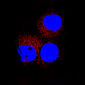
- CD155/PVR in U937 human histiocytic lymphoma cell line. CD155/PVR was detected in immersion fixed U937 human histiocytic lymphoma cell line using Goat Anti-Human CD155/PVR Antigen Affinity-purified Polyclonal Antibody (Catalog # AF2530) at 15 µg/mL for 3 hours at room temperature. Cells were stained using the NorthernLights™ 557-conjugated Anti-Goat IgG Secondary Antibody (red; Catalog # NL001) and counterstained with DAPI (blue). Specific staining was localized to cytoplasm. View our protocol for Fluorescent ICC Staining of Non-adherent Cells.