Explore
 Validate
 Learn
LearnNBP1-92246
antibody from Novus Biologicals
Targeting: CDHR2
FLJ20124, FLJ20383, PC-LKC, PCDH24, PCLKC
 Immunohistochemistry
Antibody data
- Antibody Data
- Antigen structure
- References [1]
- Comments [0]
- Validations
- Immunohistochemistry [4]
Submit
Validation data
Reference
Comment
Report error
- Product number
- NBP1-92246 - Provider product page

- Provider
- Novus Biologicals
- Proper citation
- Novus Cat#NBP1-92246, RRID:AB_11003385
- Product name
- Rabbit Polyclonal PCLKC Antibody
- Antibody type
- Polyclonal
- Description
- Immunogen affinity purified. Specificity of human PCLKC antibody verified on a Protein Array containing target protein plus 383 other non-specific proteins.
- Reactivity
- Human
- Host
- Rabbit
- Isotype
- IgG
- Vial size
- 0.1 ml
- Storage
- Store at 4C short term. Aliquot and store at -20C long term. Avoid freeze-thaw cycles.
Submitted references Proteomic analysis of the enterocyte brush border.
McConnell RE, Benesh AE, Mao S, Tabb DL, Tyska MJ
American journal of physiology. Gastrointestinal and liver physiology 2011 May;300(5):G914-26
American journal of physiology. Gastrointestinal and liver physiology 2011 May;300(5):G914-26
No comments: Submit comment
Supportive validation
- Submitted by
- Novus Biologicals (provider)
- Main image
- Experimental details
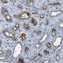
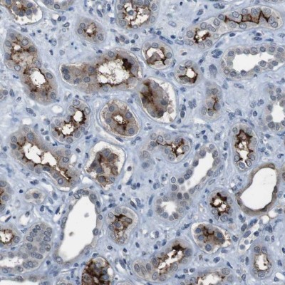
- Immunohistochemistry-Paraffin: PCLKC Antibody [NBP1-92246] - Staining of human kidney shows moderate to strong membranous positivity in cells in tubules.
- Submitted by
- Novus Biologicals (provider)
- Main image
- Experimental details
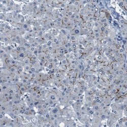
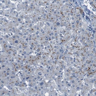
- Immunohistochemistry-Paraffin: PCLKC Antibody [NBP1-92246] - Staining of human liver shows moderate to strong membranous positivity in hepatocytes.
- Submitted by
- Novus Biologicals (provider)
- Main image

- Experimental details
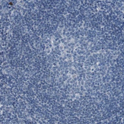
- Immunohistochemistry-Paraffin: PCLKC Antibody [NBP1-92246] - Staining of human tonsil shows no membranous positivity as expected.
- Submitted by
- Novus Biologicals (provider)
- Main image

- Experimental details
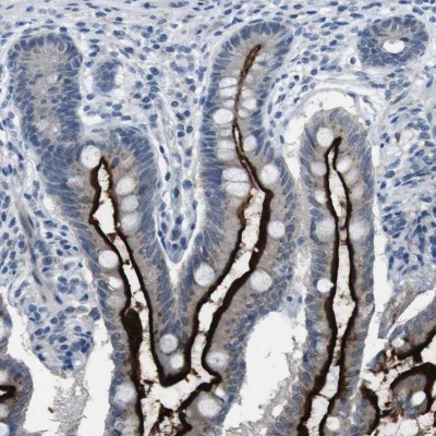
- Immunohistochemistry-Paraffin: PCLKC Antibody [NBP1-92246] - Staining of human small intestine shows positivity in apical membrane in glandular cells.