Explore
 Validate
 Learn
Learn Western blot
 Immunocytochemistry
Antibody data
- Antibody Data
- Antigen structure
- References [2]
- Comments [0]
- Validations
- Western blot [2]
- Immunohistochemistry [1]
Submit
Validation data
Reference
Comment
Report error
- Product number
- NBP1-32689 - Provider product page

- Provider
- Novus Biologicals
- Proper citation
- Novus Cat#NBP1-32689, RRID:AB_2207768
- Product name
- Rabbit Polyclonal TNIP2 Antibody
- Antibody type
- Polyclonal
- Description
- Immunogen affinity purified.
- Reactivity
- Human
- Host
- Rabbit
- Isotype
- IgG
- Vial size
- 100 ul
- Storage
- Aliquot and store at -20C or -80C. Avoid freeze-thaw cycles.
Submitted references TNIP2 is a Hub Protein in the NF-κB Network with Both Protein and RNA Mediated Interactions.
Proteins interacting with cloning scars: a source of false positive protein-protein interactions.
Banks CA, Boanca G, Lee ZT, Eubanks CG, Hattem GL, Peak A, Weems LE, Conkright JJ, Florens L, Washburn MP
Molecular & cellular proteomics : MCP 2016 Nov;15(11):3435-3449
Molecular & cellular proteomics : MCP 2016 Nov;15(11):3435-3449
Proteins interacting with cloning scars: a source of false positive protein-protein interactions.
Banks CA, Boanca G, Lee ZT, Florens L, Washburn MP
Scientific reports 2015 Feb 23;5:8530
Scientific reports 2015 Feb 23;5:8530
No comments: Submit comment
Supportive validation
- Submitted by
- Novus Biologicals (provider)
- Main image
- Experimental details
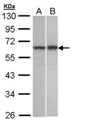
- Western Blot: TNIP2 Antibody [NBP1-32689] - Sample (30 ug of whole cell lysate)A: A431 B: Hela S310% SDS PAGE, antibody diluted at 1:1000.

- Submitted by
- Novus Biologicals (provider)
- Main image

- Experimental details
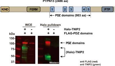
- Western Blot: TNIP2 Antibody [NBP1-32689] - A region of PTPN13 containing the PDZ domains is sufficient for association with Flexi cloned Halo-TNIP2. Part of the PTPN13 ORF coding for a 903 aa region, which included the five PDZ domains, was subcloned into FLAG-pcDNA5/FRT and coexpressed in HEK293T cells with or without Halo-TNIP2 (with the valine cloning scar) as indicated. Lysates were subjected to Halo affinity purification and the resulting eluates analysed by SDS-PAGE and Western blotting. Proteins were detected using anti-FLAG(R) M2 mouse monoclonal and anti-TNIP2 rabbit polyclonal primary antibodies, and with IRDye(R) 680LT labeled anti-mouse (red) and IRDye(R) 800CW anti-rabbit (green) secondary antibodies. Proteins were visualized using a LI-COR(R) Odyssey(R) infrared imaging system. Image collected and cropped by CiteAb from the following publication (http://www.nature.com/articles/srep08530) licensed under a CC-BY licence.
Supportive validation
- Submitted by
- Novus Biologicals (provider)
- Main image
- Experimental details
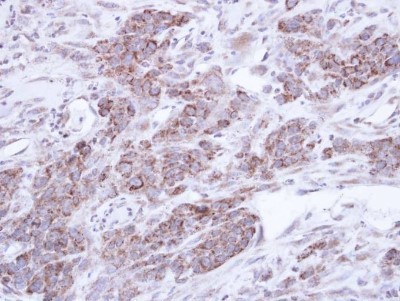
- Immunohistochemistry-Paraffin: TNIP2 Antibody [NBP1-32689] - Paraffin-embedded SAS xenograft, using antibody at 1:500 dilution.